
> New Molecule Discovery > Molecular Docking Techniques > Forge Precision: Cleaning Protein Structures for Docking Excellence
Forge Precision: Cleaning Protein Structures for Docking Excellence
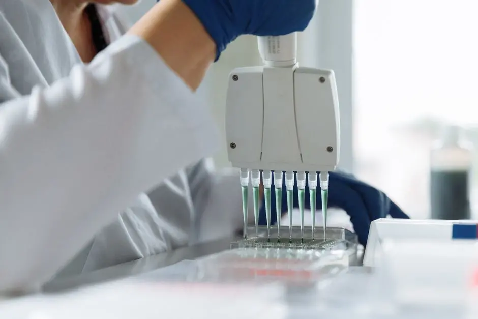
In the relentless pursuit of novel therapeutics and advanced biological insights, molecular docking stands as a cornerstone technique. Yet, its transformative power hinges on a foundational, often underestimated, prerequisite: impeccably prepared protein structures. Imagine constructing a high-performance engine with faulty blueprints; the outcome is destined for failure. Similarly, attempting molecular docking with an uncleaned, unrefined protein structure introduces noise, inaccuracies, and ultimately, misleading results that derail drug discovery efforts.
We embark on a surgical exploration, dissecting the critical steps and insider strategies required to transform raw crystallographic data into a pristine, docking-ready receptor. This isn't merely a technical guide; it's a strategic imperative for any scientist leveraging molecular modeling. We reveal why meticulous protein preparation is not a tedious chore but a critical investment, directly correlating with the reliability and predictive power of your docking experiments. Master these protocols, and you will not only enhance your understanding of the fundamental docking methodologies used to predict molecule target interactions but also significantly accelerate your journey towards biological discovery and drug development. Let's optimize every atomic detail to conquer the next frontier in molecular design.
The Imperative of Cleanliness: Why Protein Preparation isn't Optional
We initiate our conquest of molecular docking by acknowledging a fundamental truth: the integrity of our input dictates the validity of our output. Raw protein structures sourced from databases like the Protein Data Bank (PDB) are invaluable, yet they represent a snapshot of a crystalline environment, not a pristine, docking-ready receptor. Directly using these structures for docking without meticulous preparation is akin to building a skyscraper on an unstable foundation—it inevitably leads to structural weaknesses and potential collapse. The PDB is a repository of experimental data, often containing artifacts, missing information, and components irrelevant or even detrimental to a focused docking simulation.
Common issues we confront include missing atoms in side chains or even entire loops, alternate conformations for residues with high mobility, crystallographic water molecules, co-crystallized ligands, and various heteroatoms or non-standard residues. Each of these can introduce significant errors. A missing hydrogen atom can lead to incorrect charge distribution or an inability to form a crucial hydrogen bond. An unresolved loop might create an artificial barrier or an incorrect binding pocket geometry. Crystallographic waters, while sometimes mediating interactions, can frequently obscure the true binding site or introduce steric clashes if not handled judiciously. Failing to address these discrepancies compromises the protein's electrostatic potential, its steric profile, and ultimately, its interaction landscape with potential ligands. We must actively transform this experimental data into a computationally robust model, guaranteeing that our docking simulations reflect the most accurate biological reality possible.
Initial Assessment and Corrective Actions: First Line of Defense
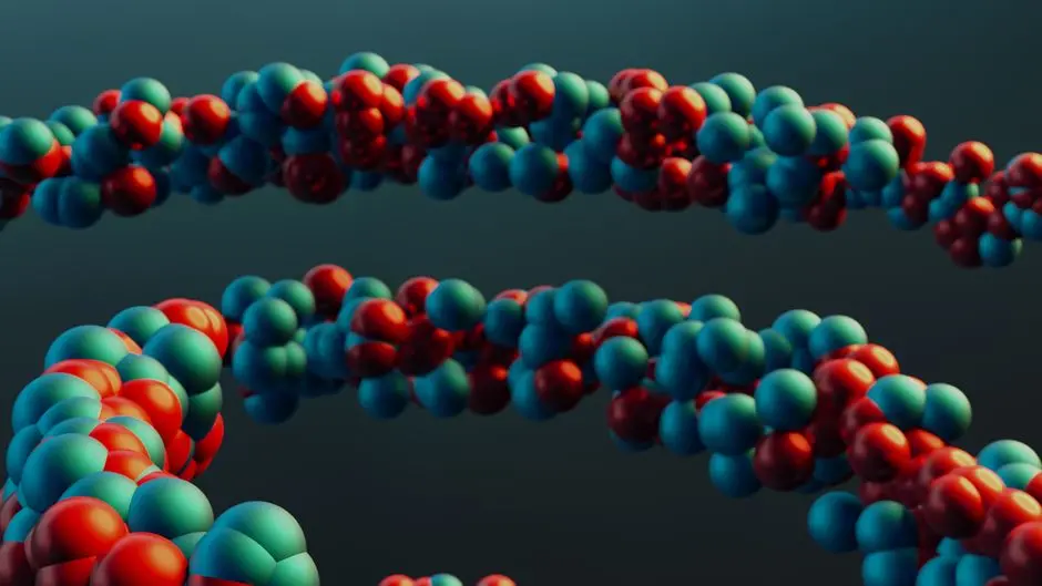
Our first strategic maneuver involves a thorough reconnaissance of the protein structure. Upon loading a PDB or mmCIF file into visualization software such as PyMOL, VMD, or Chimera, we conduct a critical visual inspection. This initial assessment is paramount for identifying macroscopic anomalies that automated tools might miss. We scrutinize the protein for large gaps indicative of missing loops or entire domains, unexpected clashes, or unusual conformations that might signal poor resolution or experimental errors. These tools empower us to view the protein's topology, identify missing backbone atoms, and detect ambiguous electron density regions.
Corrective actions often begin with addressing missing heavy atoms or entire loops. For small missing fragments, specialized software or even manual intervention might be sufficient. For larger unresolved regions, homology modeling techniques (e.g., Modeller) become indispensable, leveraging known structures of related proteins to build the missing segments. We must carefully evaluate the quality of these modeled regions, often through energy minimization and validation. Furthermore, many PDB entries show alternate conformations for certain residues, typically due to flexibility. We must select the most biologically relevant conformation or the one with the highest occupancy, ensuring a singular, unambiguous representation for docking. The goal here is to construct a complete, sterically plausible model of the protein, preparing it for the atomic-level refinements that follow. This diligent upfront work drastically reduces the propagation of errors into subsequent docking calculations.
Refining the Atomic Landscape: From Water to Protons
Having established the protein's overall structural integrity, we now execute a surgical strike on the atomic landscape, focusing on precision at the molecular level. A critical step involves the strategic removal of non-essential molecules. Crystallographic water molecules, while essential for crystal packing, often interfere with docking simulations if not located in crucial binding site positions. We typically remove most bulk water, retaining only those waters explicitly shown to mediate ligand binding through experimental evidence or highly conserved structural roles. Similarly, co-crystallized ligands, unless they are the specific target ligand for competitive studies, are removed to prepare an 'apo' protein for novel ligand screening. Ions present in the PDB file are also carefully evaluated; only those demonstrably critical for protein function or structural stability are retained.
Next, we tackle the crucial task of adding hydrogen atoms and assigning correct protonation states. PDB files often lack explicit hydrogen atoms, yet these are indispensable for accurate electrostatic interactions and hydrogen bond formation. We add hydrogens based on a chosen force field and, critically, consider the physiological pH (typically 7.4) of the biological system. Tools like PDB2PQR, H++, and PROPKA are invaluable here, predicting the protonation states of ionizable residues (e.g., histidines, aspartates, glutamates) and terminal amines/carboxylates. Incorrect protonation can lead to erroneous charge assignments and misrepresentation of binding interactions. Finally, we assign partial charges to all atoms, a step fundamental for any force field-based calculation. This is typically achieved using standard force fields (e.g., Amber, OPLS) within software packages like Antechamber. A gentle energy minimization often follows, relieving any minor steric clashes introduced by the addition of hydrogens or other modifications, ensuring a chemically sensible, low-energy conformation ready for docking.
Quality Control and Best Practices: Securing Docking Integrity
Our final phase is a rigorous quality control check, transforming our prepared protein into an unassailable receptor for docking. This step is not merely an afterthought but a critical validation of all preceding modifications. We leverage established validation tools to assess the stereochemical quality of the protein. Ramachandran plots, for instance, provide a visual representation of backbone dihedral angles, highlighting any residues in energetically unfavorable regions. Stereochemical checks ensure correct bond lengths, angles, and chirality, while clash detection algorithms identify any remaining steric overlaps that could lead to non-physical interactions during docking.
Throughout this entire process, adherence to best practices is non-negotiable. Always maintain a version control system or, at minimum, a backup of the original PDB file before any modifications. Document every step: which waters were removed, how missing loops were modeled, which protonation states were assigned, and why. This meticulous record-keeping is vital for reproducibility and troubleshooting. A common pitfall is over-minimization, which can lead to unrealistic conformations by forcing the protein into an overly relaxed state that deviates from its physiological structure. Another significant error is ignoring functionally critical water molecules or ions, mistaking them for mere crystallographic noise. We must also ensure that catalytic residues or those crucial for substrate recognition possess the correct protonation states. For output, we typically convert the cleaned protein into formats like PDBQT (for AutoDock Vina), MOL2, or a standard PDB, ensuring compatibility with our chosen docking software. Remember, protein preparation is an iterative process; sometimes, initial docking results compel us to revisit and refine our receptor model. This adaptive approach ensures maximal accuracy and unlocks the true potential of our molecular docking simulations.
Key Takeaways
Core Principles of Protein Structure Cleaning
We establish meticulous protein preparation as a non-negotiable foundation for accurate molecular docking. The process involves:
- Initial Assessment: Visually inspect and identify missing atoms, loops, or alternate conformations.
- Repair and Refinement: Model missing regions, select optimal conformations, and gently minimize to resolve clashes.
- Atomic Landscape Adjustment: Strategically remove non-essential water, heteroatoms, and co-crystallized ligands.
- Hydrogen Atom and Charge Assignment: Add hydrogens, assign correct protonation states (typically at physiological pH), and calculate partial charges.
- Quality Control: Validate stereochemistry (Ramachandran plots), detect clashes, and document all modifications for reproducibility.
Impact on Docking Accuracy
Every step in protein cleaning directly influences the reliability of docking simulations. Unresolved issues lead to:
- Inaccurate binding site representation.
- Erroneous electrostatic and steric interactions.
- False positive or false negative docking predictions.
- Misleading binding poses and affinities.
A pristine protein structure is an investment that guarantees more robust, predictive, and biologically relevant docking results, accelerating drug discovery and target validation efforts.
FAQ
-
Why can't I simply use the raw PDB file directly for molecular docking?
Raw PDB files contain experimental data that often includes missing atoms, alternate conformations, crystallographic water molecules, and co-crystallized ligands that are irrelevant for a new docking experiment. These elements introduce inaccuracies in charge distribution, steric profiles, and binding site definition, leading to unreliable docking results. Direct use compromises the integrity and predictive power of your simulations.
-
How do I decide whether to keep or remove water molecules from the binding site?
The decision is critical. Remove bulk water and those not directly mediating interactions. Retain water molecules if experimental evidence (e.g., high B-factors, conserved positions across multiple structures, structural biology literature) strongly suggests they play a crucial role in ligand binding or protein stability. Often, performing docking with and without specific waters can reveal their impact.
-
What is the biggest mistake people make during protein preparation for docking?
A common and critical error is insufficient attention to protonation states, especially for ionizable residues within or near the binding site. Incorrect protonation leads to inaccurate charge assignments, which drastically impact electrostatic interactions and hydrogen bonding patterns—fundamental drivers of ligand binding. Neglecting to add hydrogens or validate stereochemistry are also major pitfalls.
-
What pH should I use when protonating the protein structure?
We typically protonate the protein at a physiological pH, commonly 7.4, to best mimic the biological environment in which the ligand-protein interaction occurs. However, if the binding process is known to occur in a specific acidic or basic environment (e.g., endosomes), adjust the pH accordingly. Always justify your chosen pH value.