
> New Molecule Discovery > Molecular Docking Techniques > Rectify Protein Preparation Errors for Optimal Docking Success
Rectify Protein Preparation Errors for Optimal Docking Success
In the relentless pursuit of novel therapeutics, molecular docking stands as an indispensable computational tool, accelerating the identification of potential drug candidates. Yet, the robustness and reliability of docking results hinge critically on one often-underestimated factor: the meticulous preparation of the protein target. Unseen errors in this foundational step can propagate through the entire simulation, leading to misleading binding predictions, wasted resources, and stalled discovery pipelines. We are here to decode the common pitfalls that plague protein preparation, transforming potential setbacks into strategic advantages.
This deep dive equips you with the surgical precision required to transform raw structural data into a biologically relevant and docking-ready protein model. We will dissect the nuances of protonation, resolve missing structural elements, navigate the complexities of cofactors, and unveil the critical role of judicious energy minimization. Elevate your research by mastering these foundational techniques, ensuring your computational experiments yield actionable insights. Unlock the full predictive power of your simulations and redefine the accuracy of your binding predictions, whether you are exploring new compounds or refining existing ones. This expertise is crucial for accurately predicting molecular interactions and is a cornerstone in applying effective docking methods to predict molecule target interactions.
The Foundational Challenge: Understanding Protein Structure Imperfections
The journey to accurate molecular docking begins with a critical truth: raw protein structures, typically sourced from the Protein Data Bank (PDB), are inherently imperfect. These crystallographic or cryo-EM models often present a snapshot that, while invaluable, requires significant refinement before computational use. A common mistake lies in assuming a PDB file is 'docking-ready' out of the box. We must recognize the fundamental limitations: missing atoms (especially hydrogens, invisible in X-ray crystallography below ~2 Å resolution), incomplete residues or loops, non-standard residue names, and ambiguities in atom typing or bond orders. Ignoring these initial imperfections is akin to building a skyscraper on sand; the entire edifice of your docking study becomes unstable.
Our first mission is to approach each PDB entry with a 'forensic' mindset. We must systematically scrutinize the structural data for discrepancies. Common error: Proceeding without initial PDB validation. Tools like PDBFixer, MolProbity, or even basic visualization software can highlight glaring issues. We forge a robust workflow by first removing unwanted molecules (excess water, cryo-protectants unless biologically critical), correcting atom names to standard conventions, and ensuring consistent residue numbering. This initial phase, often perceived as tedious, is the bedrock of reliable docking. We convert potential liabilities into assets by establishing a clean, unambiguous starting point, ensuring all subsequent preparation steps build upon a solid, interpretable foundation.
Protonation State: The Silent Determinant of Interaction Geometry
One of the most insidious and frequently overlooked errors in protein preparation for docking is the incorrect assignment of protonation states for ionizable residues. Histidine, lysine, arginine, aspartate, and glutamate residues possess side chains that can gain or lose protons depending on the physiological pH of the system. The PDB models typically reflect experimental conditions, which may not align with the biological context (e.g., pH 7.4). Common error: Failing to adjust protonation states for biological relevance. A misprotonated residue will exhibit an incorrect charge, profoundly altering electrostatic interactions with the ligand and potentially flipping binding orientations or even preventing binding altogether.
We must surgically determine the appropriate protonation states. This involves predicting pKa values for individual residues within their specific protein environment, a task more complex than using generic pKa tables due to local electrostatic effects. Tools like PROPKA, H++ or APBS are invaluable here, as they calculate pKa values by considering the protein's 3D structure and surrounding dielectric. Once pKa values are predicted, we protonate residues according to the target pH. Insight: Pay particular attention to active site histidines; their imidazole ring can exist in various tautomeric and protonated forms (Nδ-H, Nε-H, or doubly protonated), each influencing binding differently. Optimizing these states unlocks a more accurate representation of the protein's chemical landscape, critical for precise ligand recognition and binding affinity prediction.

Missing Residues and Loop Modeling: Filling the Gaps Accurately
Incomplete protein structures are a recurring challenge, particularly when dealing with flexible regions like loops that are difficult to resolve experimentally. PDB entries often contain gaps, marked by missing backbone atoms or entire stretches of residues. Common error: Ignoring missing segments or using simplistic gap-filling methods without validation. These gaps, if unaddressed, create artificial breaks in the polypeptide chain, leading to non-physical conformations and erroneous active site geometries. Docking into a protein with a truncated binding pocket will inevitably yield irrelevant results, as critical interaction points might be absent.
We conquer this challenge through strategic loop modeling. For small gaps (1-5 residues), knowledge-based methods or energy minimization protocols can often rebuild the missing segments by sampling plausible conformations. For larger gaps, more sophisticated comparative modeling or de novo loop prediction tools become essential. The goal is not just to 'fill the gap' but to generate a conformation that is sterically plausible, energetically stable, and functionally relevant. Post-modeling, we must validate the generated loops using Ramachandran plots, clash detection, and by assessing their impact on the active site topology. This meticulous gap-filling ensures continuity and fidelity of the protein model, vital for accurate ligand positioning. We transform fragmented data into a complete, coherent biological entity ready for interaction.

Cofactors, Metal Ions, and Water Molecules: Essential but Tricky Components
Beyond the polypeptide chain, proteins often harbor non-protein entities—cofactors, metal ions, and water molecules—that are integral to their structure and function. Their presence or absence, and their precise positioning, can dramatically influence ligand binding. Common error: Indiscriminately removing cofactors or structural waters without biological justification. For instance, a metal ion in an enzyme's active site might be crucial for catalysis and ligand coordination; its removal renders the binding site artificially empty. Similarly, a highly conserved water molecule often mediates key hydrogen bonds between the protein and ligand; its arbitrary deletion disrupts critical interaction networks.
Our strategy is one of judicious preservation and informed decision-making. We must research the biological role of each non-protein component. If a cofactor is essential for the protein's activity or known to be directly involved in ligand binding, we must retain it and ensure its geometry is correct. For metal ions, ensuring their correct coordination geometry and charge state is paramount. Structural water molecules, often identified by their low B-factor and conserved hydrogen bonding networks, should be retained. Conversely, bulk solvent molecules, often numerous and disordered, are typically removed. The discerning removal versus retention of these components demands expert judgment, transforming ambiguity into functional clarity for effective docking simulations. We empower the protein model with its full biological context.

Energy Minimization and Force Fields: Over-optimizing or Under-refining
Once the protein structure is complete, protonated, and decorated with essential non-protein components, the next critical step is energy minimization. This process optimizes the geometry of the molecule to relieve steric clashes and bring it to a more stable, low-energy conformation, using a defined force field. Common error: Over-minimization, leading to an 'over-optimized' state that is no longer biologically relevant, or under-minimization, leaving residual clashes. Aggressive minimization can sometimes drive the protein into an artificial local energy minimum, distorting the active site and hindering realistic ligand binding. Conversely, insufficient minimization leaves the protein riddled with high-energy conformations that can destabilize docking poses.
We apply energy minimization with precision and restraint. We must select an appropriate force field (e.g., CHARMM, AMBER, OPLS-AA), ensuring it is compatible with both the protein and the subsequent docking algorithm. Typically, a multi-stage minimization approach is optimal: initial relaxation of side chains and hydrogen atoms, followed by constrained minimization of the backbone, and finally, a short, unconstrained minimization of the entire protein. Crucially, we prevent excessive structural drift by using positional restraints on backbone atoms or by limiting the number of minimization steps. The goal is to relieve local strain without losing the experimental structural information. A balanced minimization process primes the protein to interact realistically with the ligand, setting the stage for accurate binding prediction.
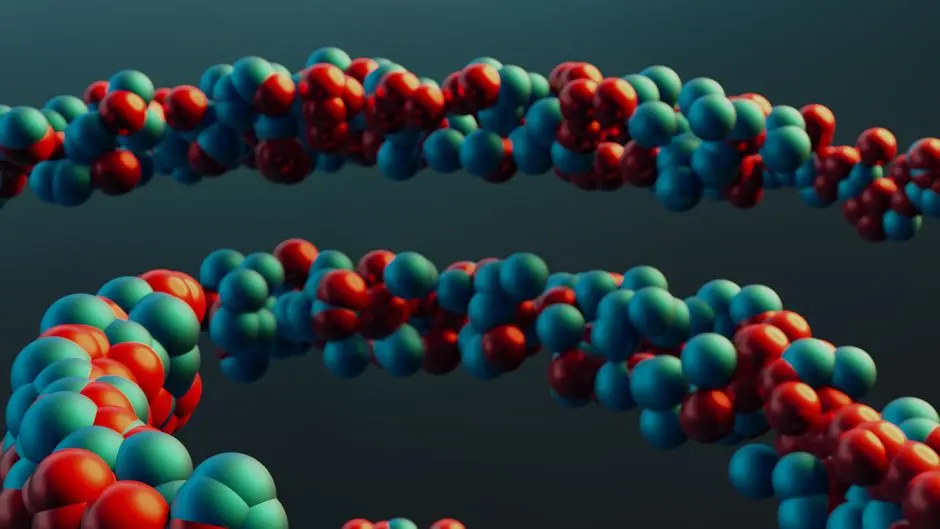
Ligand-Protein Interface: A Holistic Review of Preparation Impact
The final and perhaps most critical stage of protein preparation is a holistic review of the potential ligand-protein interface. Every error or oversight in previous steps converges here, directly impacting the accuracy of the docking process. Common error: Failing to perform a comprehensive visual and computational inspection of the active site post-preparation. A protein that looks 'fine' globally might harbor subtle but significant flaws within its binding pocket—a misaligned side chain, an incorrect charge, or an inadvertently removed critical water molecule—that will derail docking results. We must scrutinize the active site as if through a microscope, examining its electrostatics, hydrophobicity, and hydrogen bonding potential.
We conclude our preparation with a rigorous quality control. We overlay known ligands (if available) to verify that the prepared protein's active site geometry is compatible. We visually inspect for any new clashes or empty spaces introduced by our modifications. We use tools to map surface properties, ensuring that the prepared protein presents a chemically sensible environment for ligand interaction. This involves a final check of all atom types, charges, and bond orders. We empower ourselves to identify and correct any residual issues before commencing docking, ensuring the protein target is not merely clean but optimally configured to engage with potential ligands. This final, surgical review solidifies the integrity of our model, maximizing the probability of uncovering genuine biological insights from our docking simulations.
Key Takeaways
PDB Raw Data is Imperfect
Always validate PDB files first. Raw PDB structures often lack hydrogen atoms, have missing loops, or contain non-standard naming conventions. Ignoring these foundational imperfections destabilizes subsequent docking results. Utilize validation tools to identify and correct discrepancies before proceeding.
Protonation States Are Critical
Correctly assign protonation states for all ionizable residues (His, Asp, Glu, Lys, Arg). Physiological pH dictates the charge and interaction capabilities of these residues. Misassigned states lead to incorrect electrostatics, drastically altering ligand binding. Use pKa prediction tools like PROPKA to ensure biological relevance, especially for active site histidines.
Address Missing Segments Judiciously
Do not ignore missing residues or loops. Gaps create artificial breaks and inaccurate active site geometries. Employ loop modeling tools for reconstruction, ensuring steric plausibility and energetic stability. Validate rebuilt regions to maintain protein integrity and functional relevance.
Handle Non-Protein Components with Care
Retain essential cofactors, metal ions, and structural water molecules. These elements are often integral to protein function and ligand binding. Research their biological roles; remove only bulk solvent or non-essential components. Arbitrary removal can disrupt critical interaction networks and lead to false results.
Minimize Structures Precisely
Apply energy minimization with restraint using appropriate force fields. The goal is to relieve steric clashes and achieve a stable conformation without 'over-optimizing' to an artificial state. Use a multi-stage approach with positional restraints to prevent excessive structural drift, ensuring the protein model remains biologically relevant.
Conduct a Holistic Active Site Review
Perform a comprehensive post-preparation inspection of the active site. Even subtle errors can derail docking. Visually inspect electrostatics, hydrophobicity, and hydrogen bonding potential. Overlay known ligands to confirm compatibility. This final quality control is paramount for achieving genuine biological insights from docking simulations.
FAQ
-
Why can't I just use a PDB file directly for molecular docking?
PDB files, while invaluable, often contain structural imperfections such as missing hydrogen atoms (invisible to X-ray crystallography), incomplete loops, non-standard residue names, or unresolved atom positions. Directly using them can lead to incorrect charge distributions, steric clashes, and non-physiological binding site geometries, severely compromising docking accuracy.
-
How important is correct protonation state assignment in protein preparation?
It is critically important. The protonation state of ionizable residues (like His, Asp, Glu, Lys, Arg) dictates their charge and hydrogen bonding capabilities. Incorrect assignment can drastically alter the protein's electrostatic surface and active site chemistry, leading to erroneous ligand-protein interactions and misleading docking results. Tools like PROPKA help predict these states for physiological relevance.
-
Should I always remove all water molecules from the PDB structure before docking?
No, this is a common mistake. While bulk solvent water molecules are typically removed, certain 'structural' or 'bridging' water molecules within the active site can be crucial for ligand binding by mediating hydrogen bonds. These should be identified (e.g., by low B-factors or conservation) and retained if they play a significant role in the binding mechanism, as their removal can lead to false negatives in docking predictions.