
> Advanced Molecular Biology > Molecular Mechanisms of Gene Expression > Mastering Cellular Translation: Precision Protein Synthesis
Mastering Cellular Translation: Precision Protein Synthesis
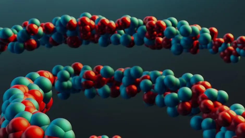
We enter the microscopic arena where life takes shape, where genetic codes transform into the functional machinery of our cells. The process of translation, this essential conversion of messenger RNA (mRNA) information into proteins, represents the beating heart of molecular biology. Without accurate and efficient translation, the cellular edifice collapses, and with it, the complexity of all life forms.
In this article, we will deconstruct the complex and fascinating molecular steps of translation. We will explore the millimeter-precise choreography of ribosomes, tRNAs, and protein factors that will orchestrate the synthesis of each protein, from initiation to termination. Understanding these mechanisms is not just an academic exercise; it is holding the key to deciphering diseases, designing new therapies, and pushing the boundaries of our knowledge about the transformation of genetic information into functional biomolecules. Let's forge a surgical understanding of this fundamental process that underpins all of modern biology. Prepare to dive into the very heart of life itself.
The Translational Apparatus: Orchestrating the Protein Factory
We delve into the heart of the process by identifying the fundamental players that make up the translation machinery. The ribosome stands as the central pivot, a complex cellular factory composed of ribosomal RNA (rRNA) and numerous proteins. In eukaryotic cells, we operate with 80S ribosomes, formed from a large 60S subunit and a small 40S subunit. Each subunit possesses distinct sites – the A (aminoacyl), P (peptidyl), and E (exit) sites – crucial for the progression of protein synthesis. This architecture ensures the protection of mRNA and the efficiency of amino acid assembly. In parallel, transfer RNAs (tRNAs) act as molecular adaptors, each carrying a specific amino acid at one end and an anticodon complementary to an mRNA codon at the other. Precise anticodon-codon pairing is the cornerstone of translation fidelity.
The accuracy of this pairing is ensured by a family of enzymes, the aminoacyl-tRNA synthetases. These enzymes charge each tRNA with the correct amino acid, a vital process for preventing errors that could lead to non-functional or toxic proteins. We also utilize mRNA as the template, its information encoded in sequences of three nucleotides, codons. The untranslated regions (UTRs) at the 5' and 3' ends of the mRNA play essential regulatory roles, influencing mRNA stability and translation efficiency. Finally, energy, primarily in the form of GTP, is consumed at each major step of translation, powering the conformational changes and movements required. These components interact with a plethora of translation factors, proteins that assist and regulate each phase, transforming the genetic code into the very substance of life.

Initiation: Launching Protein Synthesis with Precision
Translation initiation is a critical step, defining the precise start point for protein synthesis and ensuring the production of a correct protein. We trigger this process in eukaryotes by recruiting the 40S small ribosomal subunit, charged with the initiator Met-tRNA (methionine) and several eukaryotic initiation factors (eIFs), to the 5' cap of the mRNA. This 43S pre-initiation complex then scans the mRNA in a 5' > 3' direction until it identifies the AUG start codon, often nestled within a specific nucleotide context known as the Kozak sequence. This meticulous scanning ensures the correct reading frame is established from the outset; an error here would lead to an entirely different and likely non-functional protein.
Once the AUG codon is recognized, conformational changes occur, signaling the departure of certain eIFs and allowing the arrival of the 60S large ribosomal subunit. With GTP hydrolysis by eIF5B, the two subunits assemble to form the complete 80S ribosome, with the initiator Met-tRNA positioned in the P site. This initial positioning is of paramount importance: it anchors the first brick of the future protein and establishes the precise alignment for subsequent additions. A common error, faulty regulation of eIFs, can lead to initiation at non-canonical AUG codons, yielding truncated or aberrant proteins, a phenomenon observed in several human diseases, including certain cancers. We master this phase to ensure flawless protein production, an absolute requirement for cellular health.
Elongation: Building the Polypeptide Chain Amino Acid by Amino Acid
Once the initiation complex is formed, we move on to the elongation phase, where the polypeptide chain sequentially lengthens, one amino acid at a time. This cyclical process occurs on the assembled 80S ribosome and involves a precise choreography between the A, P, and E sites. The first step of each elongation cycle is the recruitment of a new aminoacyl-tRNA to the A site of the ribosome. This recruitment is orchestrated by a major elongation factor, eEF1A (eukaryotic Elongation Factor 1A), which, bound to a GTP molecule, delivers the charged tRNA to the A site. If the codon-anticodon pairing is correct, GTP is hydrolyzed, and eEF1A releases the tRNA, allowing the amino acid to be incorporated.
Next, the peptidyltransferase activity, residing in the rRNA of the large ribosomal subunit, catalyzes the formation of a peptide bond between the amino acid in the A site and the nascent polypeptide chain carried by the tRNA in the P site. The chain is thus transferred from the tRNA in the P site to the tRNA in the A site. The third step, translocation, is triggered by eEF2 (eukaryotic Elongation Factor 2), which, through GTP hydrolysis, causes the ribosome to move along the mRNA by one codon (three nucleotides). This movement shifts the uncharged tRNA from the P site to the E site, and the tRNA carrying the extended polypeptide chain from the A site to the P site. The uncharged tRNA then exits the E site, clearing the way for the next cycle. We repeat this cycle hundreds, even thousands of times, at a rate of several amino acids per second, to synthesize proteins of all sizes. The fidelity of this process is crucial; elongation errors can lead to misfolded proteins and toxic cellular aggregates.
Termination: The Precise Release of the Nascent Polypeptide
Following elongation, we reach the termination phase, a vital checkpoint for the release of the complete and functional protein. Termination is triggered when the ribosome encounters a stop codon on the mRNA. In eukaryotes, there are three stop codons: UAA, UAG, and UGA. Unlike amino acid codons, no tRNA possesses an anticodon complementary to these stop codons. Instead, release factors (eRFs for eukaryotes) recognize these specific sequences. The universal eukaryotic release factor, eRF1, is a protein that mimics the shape of a tRNA and binds to the A site when one of the stop codons is present.
Once eRF1 is bound, it interacts with another release factor, eRF3, which is a GTPase. GTP hydrolysis by eRF3 induces a conformational change that stimulates the peptidyltransferase activity of the large ribosomal subunit, but instead of forming a new peptide bond, it catalyzes the hydrolysis of the bond between the polypeptide chain in the P site and its tRNA. This cleavage releases the newly synthesized protein into the cytoplasm. Ribosomes then dissociate into their 40S and 60S subunits, which are recycled for new rounds of translation. Ribosome dissociation and recycling factors (such as ABCE1) assist this process. Premature or faulty termination can have disastrous consequences, producing truncated proteins or causing the formation of stalled ribosomes, which must be resolved by quality control mechanisms to maintain cellular homeostasis. We ensure optimal release of biomolecules with absolute precision, validating each step of their manufacturing.

Advanced Translational Control and Therapeutic Implications
Beyond the fundamental steps, we explore the sophisticated regulation of translation, a level of gene control crucial for cellular adaptation and development. Translation regulation can be global, affecting protein synthesis in general, often in response to cellular stresses such as nutrient deprivation or heat shock, or specific, targeting particular mRNAs. Elements in the 5' and 3' untranslated regions (UTRs) of mRNAs can bind proteins or microRNAs (miRNAs) that modulate initiation efficiency or mRNA stability. For example, the presence of upstream open reading frames (uORFs) upstream of the main start codon can repress the translation of the major protein by altering ribosomal scanning.
We also observe polyribosome or polysome phenomena, where multiple ribosomes simultaneously translate the same mRNA, thereby maximizing protein synthesis efficiency. This coordination is essential for rapidly responding to the cell's protein needs. Co-translational processes, such as chaperone-assisted folding and targeting of proteins to specific organelles via signal peptides, often begin even before translation is complete, optimizing protein functionality from its inception. Dysregulations of translation are implicated in major pathologies, from neurodegeneration to cancer, where aberrant protein synthesis leads to disrupted cellular functions. Drugs, such as many antibiotics that selectively target bacterial translation without affecting eukaryotic translation, already exploit these differences. We are thus forging promising therapeutic avenues by refining our understanding of the precise mechanisms of translation control, opening pathways to manipulate protein production to our advantage.
Key Takeaways
The Core Translational Machinery
Translation relies on a complex interplay of ribosomes (80S in eukaryotes), tRNAs charged with specific amino acids by aminoacyl-tRNA synthetases, and mRNA carrying the genetic code. Energy in the form of GTP fuels conformational changes, while various translation factors orchestrate each step. Understanding these components is fundamental to grasping protein synthesis.
Initiation: The Precise Starting Point
Initiation sets the reading frame. The 40S ribosomal subunit, initiator Met-tRNA, and eIFs bind the 5' cap, scan for the Kozak-embedded AUG start codon, and then recruit the 60S subunit to form the functional 80S ribosome, positioning the initiator tRNA in the P site. This ensures accurate protein synthesis from the very first amino acid.
Elongation: Building the Chain
Elongation proceeds in cycles: aminoacyl-tRNA delivery to the A site (via eEF1A), peptide bond formation (peptidyl transferase activity of rRNA) linking the new amino acid to the growing chain, and translocation (via eEF2) moving the ribosome along the mRNA. This iterative process precisely adds amino acids, extending the polypeptide.
Termination: Releasing the Functional Protein
Translation halts when a stop codon (UAA, UAG, UGA) enters the A site. Release factors (e.g., eRF1) recognize these codons, triggering the hydrolysis of the bond between the polypeptide and the P-site tRNA. This liberates the complete protein, and the ribosomal subunits dissociate for recycling.
Advanced Control & Clinical Relevance
Translational regulation is highly sophisticated, influenced by mRNA UTRs, uORFs, and microRNAs. Co-translational processes like folding and targeting begin early. Dysregulation is linked to numerous diseases, and the unique mechanisms of translation are exploited in therapeutic strategies, such as antibiotics that selectively target bacterial ribosomes.
FAQ
-
What is the primary role of tRNA in translation?
tRNA (transfer RNA) acts as a molecular adaptor. It carries a specific amino acid at one end and has an anticodon at the other end. This anticodon precisely base-pairs with a complementary codon on the mRNA, ensuring that the correct amino acid is incorporated into the growing polypeptide chain according to the genetic code.
-
How do eukaryotic ribosomes locate the correct start codon on mRNA?
Eukaryotic ribosomes utilize a scanning mechanism. The 40S ribosomal subunit, along with the initiator tRNA and eIFs, binds to the 5' cap of the mRNA and scans along the mRNA in the 5' to 3' direction until it encounters the first AUG codon, often embedded within a specific sequence context called the Kozak sequence, which signals it as the authentic start codon.
-
What is the function of release factors during translation termination?
Release factors (eRFs in eukaryotes) are proteins that recognize stop codons (UAA, UAG, UGA) in the A site of the ribosome. Instead of bringing an aminoacyl-tRNA, they facilitate the hydrolysis of the bond between the polypeptide chain and the tRNA in the P site, leading to the release of the newly synthesized protein and the subsequent dissociation of the ribosomal subunits.