
> New Molecule Discovery > Biological Testing of Candidate Molecules > Deciphering Cell Assay Results: Overcoming Interpretation Hurdles
Deciphering Cell Assay Results: Overcoming Interpretation Hurdles
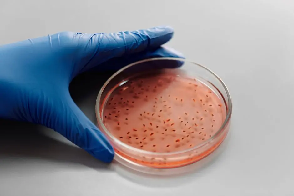
We are forging the future of molecule discovery, and in this exciting quest, the interpretation of cell assay results stands as a central, often underestimated challenge. Every data point, every activity curve, is a potential window into a new therapy, but the glass is rarely clear. The inherent complexity of in vitro biological systems, coupled with experimental variability, can obscure the pharmacological truth or, worse, lead us astray.
This article is not a mere exposition; it is a surgical action plan to outwit common pitfalls, optimize our analysis strategies, and extract maximum value from every experiment. We will unveil the subtleties, hidden biases, and best practices for transforming raw data into clinically relevant insights. Crucially, robustness for the experimental validation of bioactive compounds is paramount for navigating these complex waters. Prepare to sharpen your critical eye and turn your interpretation challenges into levers of success for novel molecule discovery.

Mastering the Intrinsic Variability of Cellular Systems
The first line of defense against misinterpretations lies in a thorough understanding of the inherent variability in cellular models. We must recognize that our cells are not static, uniform entities; they are dynamic, heterogeneous, and influenced by a multitude of factors. Cell passage variability is a frequent culprit: cell lines maintained in culture for many passages can alter their phenotypes, responses, and even their genomes. Rigorous passage monitoring and the use of low-passage cell stocks are imperative. Similarly, mycoplasma contamination, often undetectable visually, can profoundly alter cellular metabolism and signaling, skewing all results. Regular testing and strict decontamination procedures constitute essential surgical practice.
Beyond the cells themselves, the culture environment is a major source of noise. Reagent quality, particularly lots of fetal bovine serum (FBS), media composition, or even ultra-pure water, can vary and impact cell growth and responsiveness. We must systematically validate each new lot of critical reagent before its integration. Inter-operator variability, while often underestimated, can introduce significant biases. Rigorous protocol standardization, continuous training, and internal audits drive increased reproducibility. We must also consider subtle environmental factors such as temperature gradients in incubators or vibrations, which, while minor, can influence cellular physiology and our results.

Overcoming Technical Pitfalls and Assay Artifacts
Beyond biological variability, a multitude of technical pitfalls can induce artifacts, leading to erroneous conclusions. The quality and stability of the tested compounds are fundamental. A poorly soluble, unstable in solution, or aggregating compound may exhibit apparent activity that is not pharmacological. We must evaluate the solubility, stability, and aggregation state of our candidate molecules under assay conditions. Detection techniques, particularly fluorescence or luminescence measurements, are also subject to interference. Some compounds can be autofluorescent or chemiluminescent, producing false-positive signals. The inclusion of appropriate controls – cells without compound, compound without cells, or even buffers without reagents – is a proactive strategy to identify these interferences.
The edge effect in culture plates is a classic phenomenon where peripheral wells behave differently from central wells, often due to evaporation or temperature gradients. Intelligent plate design, with unused peripheral wells or wells dedicated to controls, minimizes this impact. The choice of concentrations for dose-response curves is also critical. Overly high concentrations can mask specificity or introduce irrelevant cytotoxicity, while concentrations that are too low may miss real activity. We must rely on preliminary PK/PD data and extend our concentration ranges beyond expected values to capture the full dynamics. Finally, non-specific cytotoxicity is a factor never to be overlooked. Any apparent effect must be correlated with cell viability assays to distinguish pharmacological activity from simple cell death. Each step of experimentation is a biological opportunity that we must scrutinize with surgical rigor.

Navigating Biological Complexities and Model Relevance
The correct interpretation of cell assay results requires a rigorous assessment of the biological relevance of the model. We often operate with simplified systems that do not fully reflect in vivo complexity. 2D monolayer models, while easy to handle, lack the tissue architecture, cell-matrix interactions, and nutrient/oxygen gradients present in living organisms. The adoption of 3D models (spheroids, organoids) or co-cultures can significantly improve physiological relevance, but these models introduce their own technical and analytical challenges. We must constantly question whether our cell model adequately represents the disease or biological process we are targeting. For instance, an immortalized cancer cell line may not behave in the same way as a primary tumor.
Compensatory mechanisms and feedback loops are ubiquitous in cell biology. A molecule targeting a specific pathway may trigger compensatory activation of a parallel pathway, masking its true effect or altering the expected phenotype. Understanding the signaling network is essential to anticipate these adaptive responses. Furthermore, cellular heterogeneity within a population, even a monoclonal line, can lead to variable responses. Certain subclones may be more sensitive or resistant, diluting the observed average effect. Off-target effects are another crucial complexity: a molecule is not always as specific as we desire, and its activity may be due to an unexpected interaction with a non-primary target. Counter-target screens or orthogonal phenotypic assays are essential to uncover these effects. We must always ask ourselves: is our observation an artifact of the system or a true biological interaction, and if so, with what physiological relevance?

Optimize Data Analysis and Validation Strategy
The data analysis and interpretation phase is the culmination of our work, but it is also the most vulnerable to bias and errors if we are not rigorous. Statistical significance is just a starting point; we must evaluate the magnitude of the effect and its biological relevance. A statistically significant but minimal effect may not be translational. We use robust statistical tests, but we are wary of p-hacking and uncorrected multiple comparisons. Dose-response curve modeling requires particular attention: the choice of the fitting function (e.g., Hill, 4-parameter logistic) must be justified by the underlying biology and data quality. Poor modeling can lead to erroneous IC50/EC50 values. Confirmation bias is a silent threat. We naturally tend to give more weight to data that confirm our hypotheses. A blinded approach to analysis, where possible, and an open discussion of results within a multidisciplinary team trigger more objective interpretation. Validation by orthogonal assays is the cornerstone of any robust conclusion. If a molecule shows an effect in one assay, we confirm this activity via a different detection mechanism or a distinct cell model. For example, an enzyme activity measured biochemically should be corroborated by a cellular phenotypic assay. We must also anticipate false positives and false negatives by incorporating clear positive and negative controls with known response ranges. Every unexpected result is an opportunity to refine our understanding, not a failure to ignore. We optimize our analysis processes to forge solid decisions and accelerate molecule discovery.
Key Takeaways
Mastering Biological Variability
Cell line variability, mycoplasma contamination, and environmental influences (reagents, operators) are major sources of error. We must implement rigorous SOPs, regular quality testing, and continuous training to ensure the reproducibility and reliability of cellular data.
Technical Artifact Identification
Solubility/stability issues of compounds, assay interferences (autofluorescence), plate edge effects, and non-specific cytotoxicity can mask real activity. Smart assay design with appropriate controls and early evaluation of the physicochemical properties of molecules are crucial.
Model Relevance Evaluation
The limitations of 2D models, cellular heterogeneity, compensatory mechanisms, and "off-target" effects are biological challenges. We prioritize the use of relevant cellular models (3D, co-cultures) and understand signaling networks to anticipate complex responses and validate the specificity of our molecules.
Optimization of Analysis and Validation
Beyond statistical significance, we assess the biological relevance of the effect and utilize appropriate dose-response curve models. We combat confirmation bias through blinded analyses and systematically confirm promising findings with orthogonal trials, thereby enhancing the validity and translatability of our discoveries.
FAQ
-
How to standardize cell-based assays to minimize variability?
We implement detailed Standard Operating Procedures (SOPs), including cell line management (mycoplasma testing, limited passage number), validation of each new batch of reagent (serum, media), and regular training for all operators. The use of robotic or semi-robotic systems can also significantly reduce inter-operator variability. -
What are the warning signs for assay artifacts?
We must be vigilant for high well-to-well or plate-to-plate variability, atypical dose-response curves (e.g., U-shaped), non-specific activity in controls (solvent, vehicle), or compounds showing activity in assays not relevant to their presumed mechanism of action. Additional controls (e.g., known auto-fluorescent compounds) can help identify these interferences. -
How to validate the relevance of a cell model for screening?
We validate relevance by precisely characterizing the model (target expression, active signaling pathways, phenotypic markers of the disease). We compare its response to reference compounds (positive controls) whose effects are known in vivo. The integration of 3D models or primary cells, although more complex, also increases physiological relevance. -
What is the importance of orthogonal assays in interpreting results?
Orthogonal assays are essential for confirming the activity of a molecule via an independent detection mechanism or in a slightly different biological context. They help eliminate false positives arising from assay artifacts and strengthen confidence in the specificity and robustness of observed effects, paving the way for a deeper mechanistic understanding.