
> New Molecule Discovery > Biological Testing of Candidate Molecules > Unraveling Cell-Based Screening Assays: Core of New Molecule Discovery
Unraveling Cell-Based Screening Assays: Core of New Molecule Discovery
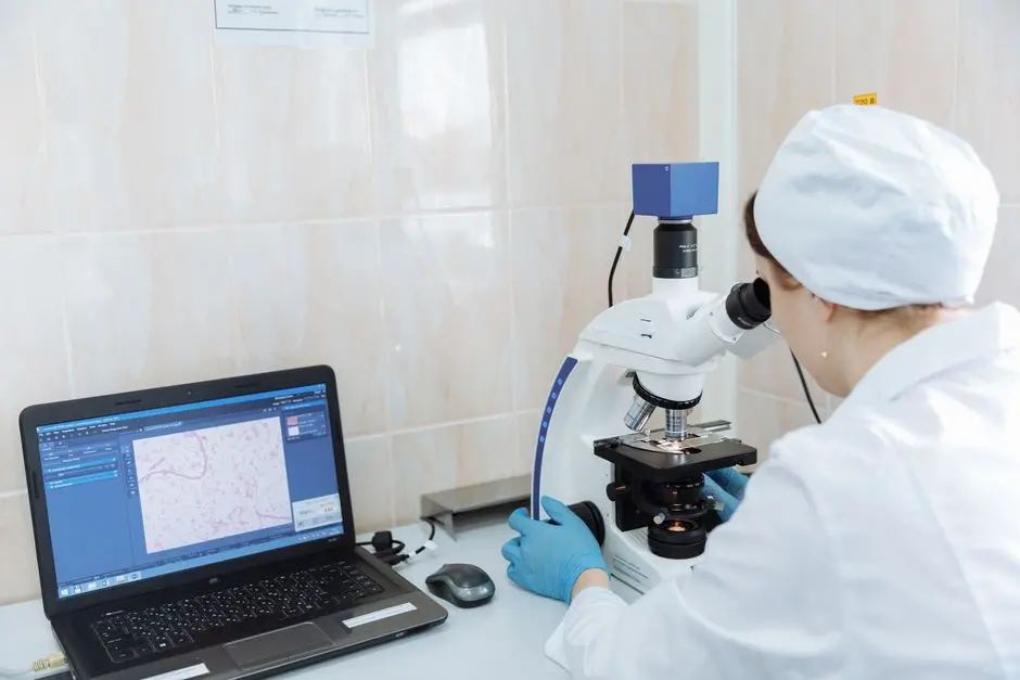
The quest for new therapeutic molecules is a grand expedition, fraught with complexity and demanding precision at every turn. How do we effectively bridge the gap from a promising chemical compound discovered in silico or through biochemical means to a potential life-saving drug? The answer lies in the strategic deployment of cell-based screening assays. These powerful tools stand as indispensable gatekeepers in the drug discovery pipeline, providing crucial insights into a molecule's biological activity, specificity, and potential toxicity within a living system.
We forge a path into understanding how these assays operate as microcosms of human biology, offering an immediate and relevant context for evaluating novel compounds. This article empowers you to master the principles, methodologies, and critical nuances of cell-based screening, transforming abstract chemical structures into agents of physiological change. We dissect the core components, from cell line selection to sophisticated readout technologies, and navigate the common pitfalls and best practices that elevate a standard experiment to a groundbreaking discovery. Prepare to revolutionize your approach to the experimental validation of bioactive compounds, ensuring that only the most promising candidates advance towards clinical development. Discover how unlocking the secrets of cell-based screening assays is not just a scientific endeavor, but a strategic imperative for shaping the future of medicine.
Foundational Principles of Cell-Based Screening Assays
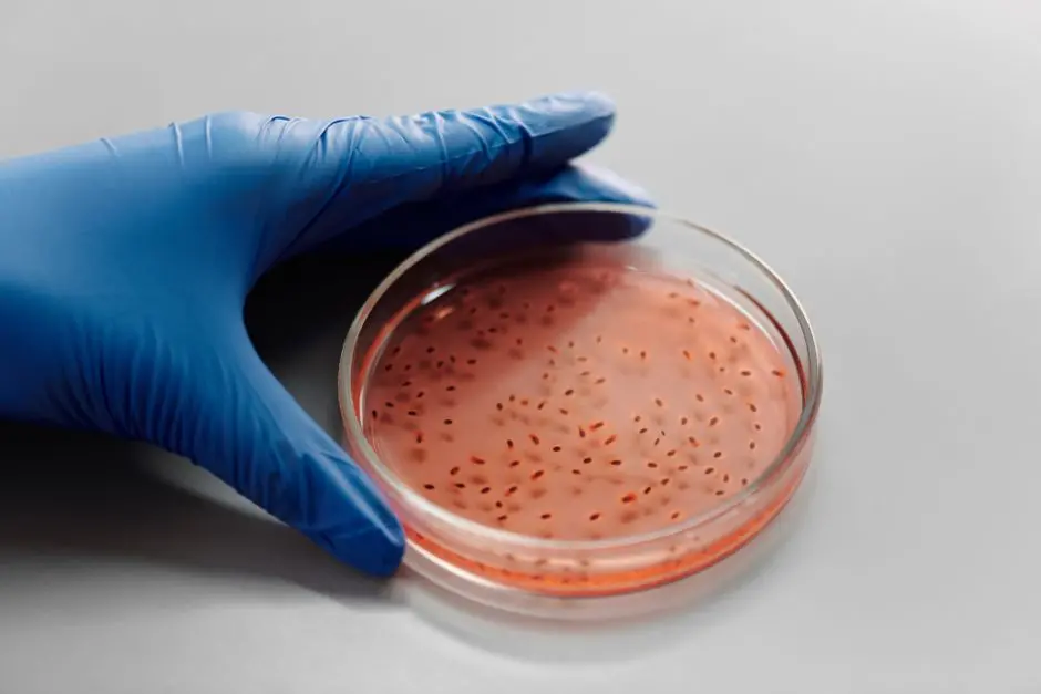
Cell-based screening assays represent a pivotal leap forward from purely biochemical or target-centric approaches in new molecule discovery. We position them as indispensable tools because they offer a more physiologically relevant environment to assess a compound's interaction with its biological target. Unlike isolated protein assays, cell-based assays encapsulate the intricate cellular machinery: membrane transport, intracellular signaling cascades, protein modifications, and compartmentalization. This holistic view is paramount for predicting in vivo efficacy and toxicity.
We classify cell-based assays primarily into two categories: phenotypic screens and target-based screens. Phenotypic screens are discovery-driven; we observe a change in cellular behavior (e.g., proliferation, morphology, migration, cell death) in response to a compound, without necessarily knowing the direct molecular target. This unbiased approach has historically led to the discovery of many first-in-class drugs. Conversely, target-based screens are hypothesis-driven. Here, we design an assay to monitor the direct engagement of a compound with a known molecular target within its cellular context, such as a specific receptor, enzyme, or signaling protein. For instance, an assay might measure the activation of a reporter gene downstream of a GPCR, confirming target engagement and functional outcome.
The immense value of cell-based screening lies in its capacity to filter out compounds that appear promising in vitro but fail to exert the desired effect in a complex cellular milieu. We leverage these assays to identify compounds that not only bind to a target but also successfully penetrate the cell membrane, remain stable within the cytoplasm, and specifically modulate the target's activity without inducing unacceptable off-target effects. This early-stage validation significantly de-risks the drug discovery pipeline, reducing the investment in molecules that are destined to fail later. We recognize that the success of these assays hinges on selecting the appropriate cell line, designing robust readouts, and rigorously controlling experimental variables. Forge ahead with us as we dismantle the complexities and construct a clear understanding of these biological powerhouses.
Consider the contrast: a biochemical assay might demonstrate a compound inhibits an enzyme with nanomolar potency. However, a subsequent cell-based assay could reveal the compound fails to penetrate the cell membrane, rendering it biologically inactive. This scenario underscores the critical "biological reality check" that cell-based assays provide. We deploy them to assess a myriad of cellular processes, from gene expression modulation and protein-protein interactions to complex pathway signaling. The readouts are diverse, encompassing fluorescence, luminescence, absorbance, high-content imaging, and even electrophysiology. Our goal is always to mirror the physiological context as closely as possible, allowing us to make informed decisions about advancing or discontinuing specific chemical series. This proactive approach ensures we optimize our resources and accelerate the journey toward impactful therapies.
Engineering Robust Cell-Based Assays: Selection and Design
The bedrock of any successful cell-based screening assay is the judicious selection and meticulous management of the cellular system. We initiate this critical phase by choosing the most appropriate cell line, a decision that profoundly impacts assay relevance and reproducibility. Our options span several categories: primary cells, which are isolated directly from tissues and offer the highest physiological relevance but exhibit limited lifespan and batch-to-batch variability; immortalized cell lines, such as HEK293 or CHO cells, which provide robust growth, high reproducibility, and are amenable to genetic modification, though they may lack certain tissue-specific features; and increasingly, induced pluripotent stem cells (iPSCs) derived cells, offering patient-specific models and the potential to differentiate into various cell types, thereby enhancing disease relevance.
Once the cell line is established, we architect the assay with principles that prioritize throughput, signal reliability, and biological relevance. A crucial design element is the establishment of a clear, measurable endpoint that directly reflects the biological question. For instance, if we aim to discover inhibitors of a specific kinase, our assay endpoint might be the phosphorylation status of its downstream substrate, measured via a fluorescent immunoassay. We rigorously define the assay window, striving for a high signal-to-noise ratio, typically quantified by the Z-factor, where a value > 0.5 indicates an excellent assay suitable for high-throughput screening. We systematically optimize parameters such as cell density, incubation times, reagent concentrations, and detection methods to ensure maximal signal separation between positive and negative controls.
Automation plays a formidable role in scaling cell-based screens. We leverage sophisticated liquid handling robots, plate readers, and high-content imaging systems to process thousands, even millions, of data points with precision and speed. This industrialization demands rigorous quality control throughout the process, from cell plating consistency to reagent dispensing accuracy. Common pitfalls we actively mitigate include cell contamination (bacterial, fungal, mycoplasma), inconsistencies in cell passaging and freezing, and batch variations in media or sera. These seemingly minor variables can dramatically impact assay performance and compound activity profiles. We implement stringent protocols for aseptic technique, regular cell line authentication, and standardized reagent preparation to safeguard the integrity of our data.
Furthermore, we must always consider the potential for compound interference with the assay readout itself. Some compounds are autofluorescent or absorb light at wavelengths used for detection, leading to false positives or negatives. We routinely employ orthogonal assays and counter-screens to identify and filter out such artifactual hits. Our strategy includes performing dose-response curves for every active compound, meticulously determining its EC50 (half maximal effective concentration) or IC50 (half maximal inhibitory concentration), and assessing its cytotoxicity in parallel. This multipronged validation ensures that we advance only truly bioactive molecules, minimizing resource expenditure on spurious findings and accelerating the pipeline for genuine therapeutic candidates.
Diverse Applications and Advanced Readout Technologies
The versatility of cell-based screening assays empowers us to interrogate virtually any cellular process relevant to disease, making them indispensable across a vast spectrum of therapeutic areas. We deploy these assays to investigate G protein-coupled receptor (GPCR) activation, enzyme inhibition, ion channel modulation, cell proliferation and apoptosis, gene expression changes, protein-protein interactions, and intricate pathway signaling. For instance, in oncology, we utilize assays measuring cell viability and proliferation to identify compounds that selectively kill cancer cells. In immunology, we monitor cytokine production or immune cell activation to find immunomodulatory agents. The beauty lies in our ability to adapt the cellular context and readout to precisely match the biological question at hand.
A critical aspect of assay design is the selection of the appropriate readout technology, which directly dictates the type of signal detected and how it's quantified. We predominantly employ label-based detection methods due to their sensitivity and adaptability. Fluorescence-based readouts are incredibly common, leveraging fluorescent probes that change intensity or wavelength upon target engagement or cellular event. Examples include FRET (Förster Resonance Energy Transfer) and BRET (Bioluminescence Resonance Energy Transfer) assays, which detect molecular proximity, or simple fluorescent dyes that quantify calcium flux or membrane potential. Luminescence-based readouts, often employing luciferase reporter genes, provide high sensitivity and a broad dynamic range, making them ideal for monitoring gene expression or pathway activation. Absorbance-based readouts, while generally less sensitive, are cost-effective and suitable for high-throughput quantification of cell viability (e.g., MTT assays) or enzymatic activity producing colored products.
Beyond bulk measurements, high-content imaging (HCI) stands as a transformative technology. We harness HCI to capture detailed morphological changes, subcellular localization of proteins, and cell-level events (e.g., neurite outgrowth, organelle integrity) across thousands of individual cells per well. This allows for multiparametric analysis, moving beyond a single endpoint to generate rich phenotypic profiles. For example, an HCI screen could identify compounds that disrupt mitochondrial networks in cancer cells, providing a deeper understanding of their mechanism of action than a simple viability assay. The data generated from HCI is complex, requiring advanced bioinformatics and machine learning algorithms for interpretation, but its informational density is unparalleled.
We continuously push the boundaries of these applications. Consider drug discovery for neurodegenerative diseases: we now employ iPSC-derived neurons and glial cells in complex co-culture systems to mimic disease pathology more accurately, using high-content imaging to track synapse formation or protein aggregation. For infectious diseases, we design assays to monitor viral replication within host cells or bacterial biofilm formation. Our rigorous selection of readout technologies, coupled with tailored cellular models, ensures we extract maximum biological intelligence from every screen, guiding us to truly impactful therapeutic candidates with unparalleled precision and foresight.

Optimizing Performance and Navigating Future Horizons
Achieving reliable and actionable results from cell-based screening assays necessitates a rigorous approach to data analysis, quality control, and continuous optimization. We assert that every active compound identified demands a thorough dose-response characterization to precisely quantify its potency and efficacy. This involves testing compounds across a range of concentrations to generate dose-response curves, from which we accurately derive key pharmacological parameters like EC50 (half maximal effective concentration) for agonists or IC50 (half maximal inhibitory concentration) for antagonists. Accurate curve fitting, often employing four-parameter logistic models, is paramount to extracting meaningful data and minimizing false positives or negatives. We meticulously evaluate curve shape, ensuring a clear sigmoidal response that signifies specific biological interaction rather than assay artifact.
Quality control (QC) is not a mere formality; it is the bedrock of assay integrity. We systematically implement metrics such as the Z-factor and signal window to gauge assay quality. A Z-factor value of >0.5 generally indicates an excellent assay with good separation between positive and negative controls, making it suitable for high-throughput screening. We monitor these parameters plate-by-plate and screen-by-screen to detect drifts in performance and trigger investigations into potential issues, such as reagent degradation or instrument malfunction. Furthermore, we include multiple internal controls on every plate: positive controls (known active compounds), negative controls (vehicle only), and often reference compounds to track assay variability over time. This meticulous QC strategy ensures that our data is robust, reproducible, and trustworthy, forming a solid foundation for critical Go/No-Go decisions.
As we optimize, we also look to the future, where advancements promise even greater physiological relevance. The transition from 2D monolayer cell cultures to 3D cell models, including spheroids, organoids, and tissue-on-a-chip systems, represents a significant leap. These 3D structures better mimic the complex cellular architecture, cell-cell interactions, and extracellular matrix environment found in vivo, leading to more predictive drug responses. For instance, cancer spheroids can recapitulate tumor microenvironments and drug penetration challenges more accurately than 2D cultures, providing a more realistic platform for anti-cancer drug screening. We are actively integrating these advanced models to reduce the attrition rate of drug candidates in later development stages.
Moreover, the convergence of cell-based assays with cutting-edge technologies like CRISPR/Cas9 gene editing and advanced bioinformatics is revolutionizing drug discovery. We leverage CRISPR to engineer precise disease models directly within target cells, creating cells with specific genetic mutations or reporter constructs that illuminate pathway activity. Combined with machine learning for data analysis, we can identify subtle phenotypic changes and predict drug mechanisms with unprecedented accuracy. We champion a proactive, iterative optimization cycle, where continuous refinement of our assays, informed by biological insight and technological innovation, propels us towards uncovering truly transformative molecules, cementing our role at the forefront of biological exploration and therapeutic advancement.
Key Takeaways
Core Value & Biological Relevance
Cell-based screening assays are paramount in new molecule discovery, offering a physiologically relevant platform to evaluate compound activity within a living cellular context. They bridge the gap between in silico/biochemical data and in vivo outcomes by accounting for cellular complexities like membrane permeability, metabolic stability, and intricate signaling pathways.
Strategic Design & Quality Assurance
Successful cell-based assays hinge on meticulous design, including appropriate cell line selection (primary, immortalized, iPSC-derived), robust assay parameters, and clear, measurable endpoints. Rigorous quality control, employing metrics like the Z-factor (>0.5 for excellent assays), and continuous optimization are critical for ensuring data integrity and reproducibility.
Advanced Applications & Future Trajectories
These assays boast diverse applications, from GPCR activation to high-content imaging for phenotypic profiling. Future developments are integrating advanced models like 3D cell cultures (spheroids, organoids) and cutting-edge technologies such as CRISPR/Cas9 editing to create more predictive and complex disease models, accelerating the discovery of transformative therapeutics.
FAQ
-
What distinguishes cell-based assays from biochemical assays?
Cell-based assays evaluate compounds within a living cellular context, reflecting complex biological interactions, membrane permeability, and metabolic stability. Biochemical assays typically involve isolated components (e.g., purified enzymes or receptors) in an artificial environment, focusing on direct molecular interaction without cellular complexities. -
What is the significance of a good Z-factor in cell-based screening?
The Z-factor is a statistical measure quantifying the robustness of an assay, indicating how well the signal for active compounds (positive control) is separated from inactive compounds (negative control). A Z-factor > 0.5 is considered excellent, signifying a reliable assay suitable for high-throughput screening with a low false-positive/negative rate. -
How do 3D cell models enhance the predictiveness of cell-based assays?
3D cell models (spheroids, organoids) recapitulate the in vivo cellular environment more closely than traditional 2D cultures, including cell-cell interactions, extracellular matrix effects, and oxygen/nutrient gradients. This leads to more physiologically relevant drug responses, improved prediction of efficacy and toxicity, and reduced attrition rates in preclinical development.