
> New Molecule Discovery > Biological Testing of Candidate Molecules > Forge Predictive Cell Assays: A Strategic Blueprint for Molecule Testing
Forge Predictive Cell Assays: A Strategic Blueprint for Molecule Testing
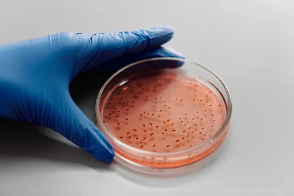
The quest to uncover novel therapeutics hinges on our ability to precisely evaluate candidate molecules, transforming abstract chemical structures into tangible biological insights. At the forefront of this intricate journey, cell assays emerge as indispensable tools – microscopic arenas where potential drugs confront their first biological challenges. We empower researchers to accelerate drug discovery by meticulously crafting these cellular battlegrounds, extracting profound data that steers the entire development process. This article unveils the critical strategies for designing potent, predictive, and high-fidelity cell assays, laying the bedrock for impactful therapeutic advancements. We dissect the nuances of selecting optimal cell models, engineering sensitive detection methodologies, and establishing robust controls that guarantee data integrity and reproducibility. Prepare to master the art and science of cell assay design, a foundational expertise that directly influences the efficacy and safety of future medicines. This mastery is not merely a technical skill; it is the strategic imperative for anyone committed to advancing the experimental validation of bioactive compounds, the cornerstone of modern pharmaceutical innovation. Join us to transform challenges into triumphs within the molecular frontier.
Foundations of Cell Assay Design: Unveiling Biological Efficacy
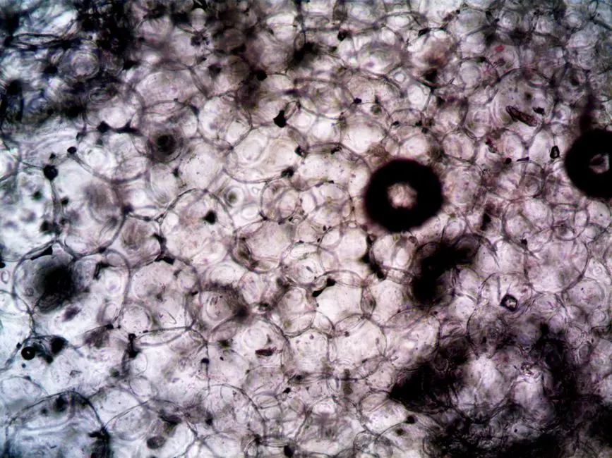
In the relentless pursuit of new therapeutic molecules, cell assays stand as the initial, critical gatekeepers, providing an indispensable bridge between abstract chemical structures and their tangible impact on living systems. We recognize that understanding a molecule's true potential demands more than isolated biochemical interactions; it requires observation within the complex, dynamic environment of a living cell. This inherent complexity grants cell assays a profound advantage over purely biochemical methods: they allow us to discern not just direct target engagement, but also intricate downstream signaling cascades, pleiotropic phenotypic alterations, and crucial off-target effects. These holistic insights are vital, often unattainable through reductionist approaches, and offer a more predictive window into a molecule’s potential in vivo behavior.
Our strategic approach to cell assay design commences with an unwavering focus on the biological question at hand. We must articulate with surgical precision: Is the objective to identify inhibitors of a specific protein pathway, activators of a receptor, or compounds that induce a desired cellular phenotype, such as differentiation, proliferation, or apoptosis? The clarity of this question dictates every subsequent design choice, from cell model selection to readout methodology. We adopt an iterative cycle of design, execution, rigorous analysis, and continuous refinement, a process that optimizes predictive power. Fundamentally, we must select appropriate cell models that faithfully recapitulate the disease state or biological process of interest, ensuring their relevance and responsiveness. Concurrently, we identify sensitive, quantifiable readouts that directly reflect the molecule's activity, establishing a clear link between stimulus and measurable response. Parameters such as cell culture conditions, media composition, cell density, and the precise duration of molecule exposure are critical and must be meticulously optimized. Forging robust cell assays is not merely a technical task; it is an act of advanced biological engineering, crafted to extract precise, actionable data that propels drug discovery forward, ultimately unlocking profound biological efficacy.
Strategic Cell Line Selection & Robust Control Engineering
The bedrock of a predictive cell assay lies in the judicious selection and rigorous management of the cellular model. We confront a spectrum of choices: primary cells, isolated directly from tissues, offer unparalleled physiological relevance but present challenges like finite lifespan and batch-to-batch variability. In contrast, immortalized cell lines, such as HEK293 or HeLa cells, provide an endless supply of homogeneous cells, ideal for large-scale screening due but carry risks of genetic drift and deviation from normal physiology. We navigate this critical decision by aligning the cell model with the specific biological question and the discovery stage. The advent of induced pluripotent stem cells (iPSCs) and their differentiated derivatives, along with patient-derived cells, further enhances translational relevance by offering disease-specific genotypes and phenotypes.
Regardless of the cell source, rigorous cell characterization is non-negotiable. We demand authentication through methods like STR profiling to prevent misidentification, and regular mycoplasma testing to ensure culture health. Maintaining consistent passage numbers is also crucial, as cellular phenotypes can subtly shift over time. Beyond cell selection, the engineering of robust controls is paramount to validate assay performance and ensure data integrity. We meticulously design: positive controls (molecules with known activity), confirming the assay’s ability to detect a response; negative controls (inactive molecules or vehicle only), establishing baseline signal and specificity; and vehicle controls (solvent used to dissolve the test molecule), accounting for any solvent-induced effects. These controls define the assay’s dynamic range and signal-to-noise ratio. By generating comprehensive dose-response curves for candidate molecules and reference compounds, we accurately determine key pharmacological parameters such as EC50 or IC50, quantifying potency and enabling direct comparison. This precision in cell model management and control engineering unlocks the true interpretive power of our assays.
Mastering Detection Methodologies & Data Readouts
The power of a cell assay culminates in its ability to translate a cellular event into a quantifiable signal. We meticulously select detection methodologies that are not only sensitive and robust but also align precisely with the biological phenomenon under investigation. A diverse arsenal of technologies empowers us to interrogate various aspects of cellular function.
Reporter gene assays, harnessing enzymes like luciferase or fluorescent proteins such as GFP, offer an elegant approach to monitor gene expression, transcription factor activity, or protein-protein interactions. They provide a highly sensitive, often high-throughput readout of pathway engagement, though we acknowledge potential limitations like transient expression variability. For assessing fundamental cellular health and proliferation, we deploy cell viability and proliferation assays. Techniques like MTT, MTS, or resazurin reduction measure metabolic activity, while ATP-based assays quantify viable cell count. BrdU incorporation directly assesses DNA synthesis. These methods provide a critical first pass for cytotoxicity and proliferative effects, guiding subsequent, more targeted investigations.
To unravel complex cellular morphology and spatial dynamics, high-content imaging emerges as an invaluable tool. By combining automated microscopy with sophisticated image analysis software, we can multiplex readouts, simultaneously assessing cell number, morphology, subcellular localization of proteins, and various phenotypic changes within individual cells or populations. This quantitative cellular profiling yields rich, multidimensional data. When focusing on specific cell populations or intricate surface marker expression, flow cytometry becomes indispensable. It allows for rapid, multiparametric analysis of thousands of cells, differentiating between cell types, quantifying protein expression, and assessing cell cycle phases or apoptosis markers with high precision. For deep dives into specific protein or gene expression patterns, we leverage techniques such as ELISA for secreted proteins, Western Blot for intracellular protein levels, and quantitative PCR for gene expression. We strategically choose our readout based on the desired level of detail, required sensitivity, dynamic range of the signal, and throughput demands. Mastering these methodologies transforms raw biological events into actionable data, providing the crucial evidence needed to advance promising molecules.
Scaling & Sophistication: High-Throughput & Advanced Models
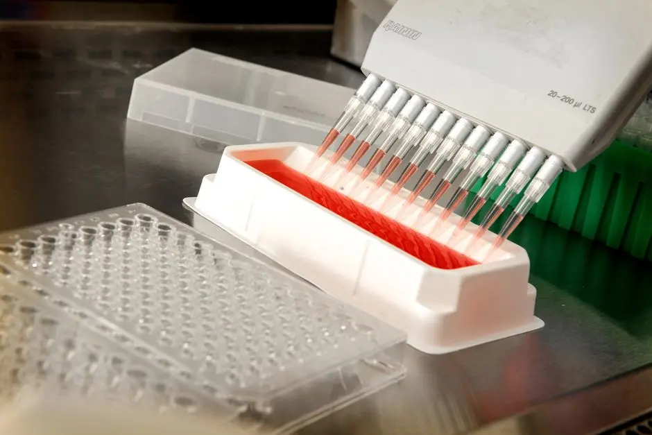
As promising molecules emerge from initial screens, the imperative shifts towards scaling assays for efficiency and enhancing their physiological relevance. We strategically embrace high-throughput screening (HTS) methodologies, transforming manual processes into automated, miniaturized operations capable of evaluating hundreds of thousands of compounds. This transition demands meticulous planning in plate formats, liquid handling robotics, and sophisticated data acquisition systems. We ensure that miniaturization does not compromise assay performance, rigorously validating Z'-factor and signal consistency across varying scales. Automated platforms enable us to rapidly identify "hits" from vast chemical libraries, accelerating the initial phase of drug discovery.
Beyond throughput, we actively integrate advanced cell models to bridge the gap between simplified 2D cell cultures and complex in vivo physiology. 3D cell cultures, including spheroids and organoids, represent a paradigm shift. Spheroids, self-assembling aggregates of cells, mimic aspects of tissue architecture and cell-cell/cell-matrix interactions more faithfully than traditional monolayers. Organoids, derived from stem cells, self-organize into miniature versions of organs with multiple cell types and functional characteristics. We leverage these models to gain more predictive insights into drug efficacy, toxicity, and penetration. While their setup and analysis demand greater expertise, their enhanced physiological relevance justifies the investment, offering a superior platform for preclinical testing.
Furthermore, we engineer co-culture systems to investigate intricate cell-cell interactions, such as those between tumor cells and immune cells. These systems illuminate how a molecule's effect on one cell type might be modulated by its microenvironment, crucial for understanding complex biological processes. The frontier of microfluidics and organ-on-a-chip technologies pushes these boundaries even further. By creating micro-environments that precisely control fluid flow and nutrient delivery, we can emulate the intricate architecture and dynamic physiological conditions of human organs. This offers an unprecedented opportunity to test drug candidates in a highly controlled, yet physiologically relevant, setting, predicting human responses with greater accuracy and reducing reliance on animal models. We champion these advanced models, understanding that their sophisticated design unlocks deeper biological truths and accelerates the translation of discovery into therapy.
Navigating Pitfalls & Forging Validation Pathways
Even the most meticulously designed cell assay can harbor subtle flaws or encounter unexpected variables. We must proactively identify and navigate common pitfalls to ensure the integrity and reliability of our data. Foremost among these are issues of off-target effects and cytotoxicity. A molecule might appear active, but its effect could be non-specific, or simply toxic to the cells at the concentrations tested, leading to false positives. We combat this by implementing orthogonal assays and rigorous cytotoxicity screens early. Other insidious challenges include assay drift over time, often due to reagent degradation or subtle changes in cell passage number, and reagent variability between different lots of antibodies, media, or compounds.
Troubleshooting is an art demanding systematic investigation. We maintain comprehensive documentation of all experimental parameters, enabling us to pinpoint inconsistencies. When results deviate from expectations, we dissect the problem by systematically checking cell health, validating reagent activity, calibrating equipment, and scrutinizing experimental protocols. Optimization is an iterative process, involving titration of reagents, adjustment of incubation times, and fine-tuning cell densities to achieve optimal signal-to-noise ratios. Crucially, a designed assay must undergo stringent validation to confirm its robustness and predictive power. We quantify assay robustness using metrics like the Z'-factor, which assesses the dynamic range and variability between positive and negative controls; a Z'-factor value >0.5 indicates an excellent assay suitable for screening. We also rigorously evaluate reproducibility through inter-day and intra-day variability studies, ensuring consistent results across different experimental runs and operators. Assaying a panel of reference compounds with known activities helps establish the assay's specificity and sensitivity, confirming it responds appropriately to relevant pharmacological agents.
Finally, we understand that robust data normalization and statistical analysis are indispensable. Raw data often requires normalization against controls to account for experimental variability. We employ appropriate statistical tests to determine significance and confidently interpret findings. Our commitment to best practices includes implementing randomized plate layouts, blinding experimental conditions to minimize bias, and maintaining an unwavering focus on detailed documentation. Each assay design, optimization, and validation step strengthens the foundation of our drug discovery efforts. By forging these rigorous validation pathways, we elevate our assays from mere experiments to powerful, predictive tools, ensuring every molecule we advance carries the stamp of scientific certainty and biological promise.
Key Takeaways
Strategic Imperatives in Cell Assay Design
We define success by aligning assay design with precise biological questions, meticulously selecting cell models (primary, immortalized, iPSC-derived) and rigorously characterizing them. Robust controls (positive, negative, vehicle) are non-negotiable for validating data integrity and quantifying molecular potency via EC50/IC50.
Advancing Detection & Predictive Models
We leverage diverse detection methodologies—reporter genes, viability assays, high-content imaging, flow cytometry, ELISA, PCR—choosing based on sensitivity and biological relevance. We scale discovery with HTS and enhance physiological fidelity using advanced models like 3D spheroids, organoids, co-cultures, and microfluidics, pushing the boundaries of preclinical predictability.
Ensuring Assay Reliability & Validation
We proactively navigate pitfalls like off-target effects and assay drift through systematic troubleshooting and optimization. Validation is critical, quantified by Z'-factor (>0.5 for excellent assays), reproducibility, specificity, and sensitivity. Rigorous data normalization, statistical analysis, and best practices (randomization, blinding, documentation) forge powerful, predictive assays.
FAQ
-
How does selecting between primary cells and immortalized cell lines impact assay predictability?
Primary cells offer higher physiological relevance, mirroring the natural tissue environment, which can lead to more predictive results for in vivo efficacy or toxicity. However, their variability and limited lifespan challenge high-throughput screening. Immortalized lines provide homogeneity and scalability, essential for early-stage discovery, but may deviate from true physiological responses due to their transformed nature. The choice balances throughput needs with the desired biological fidelity.
-
What is the significance of the Z'-factor in cell assay validation?
The Z'-factor is a critical statistical measure used to assess the quality of a high-throughput assay. It quantifies the separation between positive and negative controls relative to the variability within those controls. A Z'-factor >0.5 indicates an excellent assay with sufficient signal window and low variability, making it suitable for robust screening. Values closer to 1 signify an ideal assay, ensuring reliable distinction between active and inactive compounds and minimizing false positives or negatives.
-
Can 3D cell cultures entirely replace animal models in preclinical testing?
While 3D cell cultures, such as spheroids and organoids, significantly enhance the physiological relevance of in vitro testing compared to 2D cultures, they cannot entirely replace animal models at this stage. 3D models better mimic tissue architecture, cell-cell interactions, and microenvironments, offering improved predictability for certain aspects like drug penetration and cellular responses. However, they still lack the complete systemic complexity, intricate organ interactions, and immune responses found in a whole organism. They serve as powerful intermediate models, reducing and refining animal studies, but are not yet a complete substitute for comprehensive in vivo validation.