
> New Molecule Discovery > Biological Testing of Candidate Molecules > Unveil Potency: Master Bioactivity Assays for New Molecule Discovery
Unveil Potency: Master Bioactivity Assays for New Molecule Discovery

We are forging the future of biology, and at the heart of this pursuit lies the ability to precisely assess the impact of new molecules on biological systems. Without a rigorous understanding of the available bioactivity assay types, our drug discovery journey would be lost in uncertainty. This article is our surgical roadmap, breaking down the essential methodologies that propel a candidate molecule from hypothesis to clinical validation.
Discover how to orchestrate a high-impact screening campaign, avoiding common pitfalls and maximizing the relevance of each data point. We explore strategies for integrating in vitro and in vivo assays, deciphering the subtle biological signals that reveal a compound's true potential. Prepare to sharpen your experimental arsenal and grasp the complexity of molecular interactions. To further our expertise in validation, we will explore the fundamental principles of experimental validation of bioactive compounds, an indispensable foundation for transforming every molecule into a tangible therapeutic promise. Let us embark on this exploration, for the precision of bioactivity assays dictates our ability to sculpt groundbreaking therapies.

Deciphering Bioactivity Assays: Foundation of Molecular Discovery
In the relentless pursuit of novel therapeutics, establishing the biological activity of newly synthesized or isolated molecules stands as our initial, non-negotiable frontier. Bioactivity assays are not merely experiments; they are the interrogations we pose to molecules, demanding answers about their potential to interact with biological systems and elicit specific effects. We leverage these assays as our primary tools to identify, characterize, and optimize lead compounds, guiding them from raw chemical entities to viable drug candidates.
Understanding the spectrum of bioactivity assays is paramount. We differentiate them broadly into two main categories: biochemical assays, which typically involve purified molecular targets in a controlled environment, and cell-based assays, which assess molecular effects within the complex milieu of living cells. Each category offers distinct advantages and limitations, compelling us to strategically select the most appropriate assay for each stage of our discovery pipeline. For instance, biochemical assays offer high precision for target engagement, while cell-based assays provide crucial insights into cellular permeability, toxicity, and overall phenotypic response.
The journey from a screening hit to a validated lead compound is iterative. We initiate with high-throughput screening (HTS) to cast a wide net, identifying compounds with preliminary activity. Subsequent validation and characterization involve a cascade of progressively more complex and physiologically relevant assays, meticulously refining our understanding of a molecule's mechanism of action, potency, and selectivity. Our ultimate goal is to generate robust, reproducible data that stands as an unshakeable testament to a molecule's therapeutic potential, propelling it towards further preclinical and clinical development. We must embrace a holistic view, integrating various assay types to paint a comprehensive picture of bioactivity.
Cell-Based Assays: Unlocking Physiological Relevance and Cellular Responses
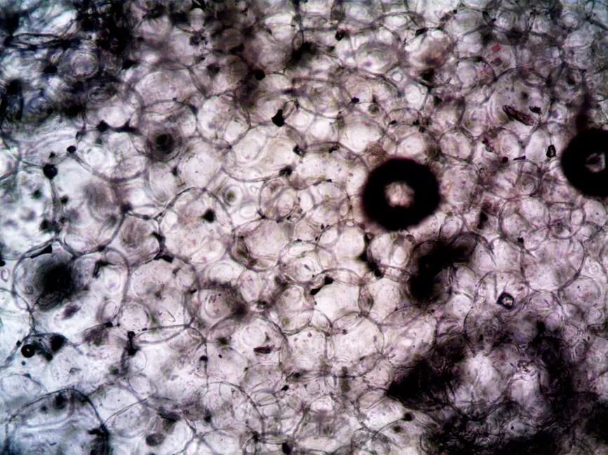
Cell-based assays are the bedrock of understanding how a molecule interacts within a living cellular context. These assays capture a molecule’s ability to permeate cell membranes, engage intracellular targets, and trigger downstream signaling pathways, providing a more physiologically relevant snapshot than isolated biochemical systems. We deploy them to assess a myriad of cellular functions, from proliferation and viability to gene expression, protein translocation, and specific pathway activation.
Consider cell viability and proliferation assays, such as MTT, MTS, or resazurin assays, which quantify metabolic activity or cell count changes, serving as fundamental indicators of cytotoxicity or growth promotion. These are crucial for initial toxicity assessments. Reporter gene assays, where a specific promoter drives the expression of a detectable protein (e.g., luciferase, GFP) in response to molecular stimulation, allow us to directly monitor gene regulation and pathway activation. These offer excellent sensitivity for transcriptional activity.
Functional assays dive deeper, measuring specific cellular responses like calcium flux (indicating ion channel or GPCR activation), receptor internalization, or the production of second messengers (e.g., cAMP, IP3). Advanced techniques like high-content imaging (HCI) further revolutionize this space, enabling multiplexed measurements of cellular morphology, protein localization, and cell cycle progression within individual cells. This capability delivers a wealth of spatial and temporal data. However, we must remain vigilant: cell line selection is critical, as genetic drift and passage number can significantly impact assay reproducibility. We constantly optimize culture conditions and ensure cell line authenticity to guarantee robust and meaningful data.

Biochemical Assays: Pinpointing Molecular Interactions with Precision
Biochemical assays empower us to dissect molecular interactions at a fundamental level, providing direct evidence of a molecule's engagement with its specific target. Unlike cell-based assays, these typically employ purified proteins, enzymes, nucleic acids, or lipids in an cell-free environment. This reductionist approach eliminates cellular complexities, offering unparalleled precision in measuring binding affinities, enzyme kinetics, and direct modulation of target activity. We initiate these assays when a specific molecular target has been identified or hypothesized.
Enzyme activity assays represent a cornerstone, measuring the catalytic activity of enzymes and the inhibitory or activating effects of candidate molecules. We quantify product formation or substrate depletion over time, often using fluorescent or colorimetric readouts. Data derived from these assays, such as IC50 values, directly reflect a molecule's potency against its enzymatic target. Furthermore, we leverage protein-protein interaction (PPI) assays—like FRET (Förster Resonance Energy Transfer), BRET (Bioluminescence Resonance Energy Transfer), or AlphaLISA—to detect and quantify the binding of two proteins, and how a small molecule might modulate this interaction. This is vital for targets that rely on complex formations.
Receptor-ligand binding assays, traditionally using radioligands but increasingly employing label-free technologies like Surface Plasmon Resonance (SPR), directly quantify the affinity and kinetics of a molecule binding to its receptor. SPR provides real-time interaction data, offering insights into association and dissociation rates. While highly precise, a critical caveat for biochemical assays is their isolation from the cellular context. A molecule active in a biochemical assay might fail in cells due to poor permeability, metabolic instability, or off-target effects. We consistently integrate these data with cell-based findings to bridge the gap toward biological relevance.

Phenotypic and Ex Vivo Assays: Bridging In Vitro to In Vivo Systems
As we advance in drug discovery, moving beyond isolated molecular targets or single cell lines becomes imperative. Phenotypic and ex vivo assays represent critical intermediary steps, bridging the gap between simplified in vitro models and complex in vivo systems. These assays allow us to observe a molecule's effect on a more integrated biological process or within a more complex tissue environment, often without a predefined molecular target.
Phenotypic screening, for example, focuses on identifying molecules that induce a desired change in cell behavior or morphology, irrespective of the initial target. We might screen for compounds that restore a healthy phenotype in diseased cells, induce differentiation, or inhibit metastasis. This approach has gained resurgence, particularly for complex diseases where target identification remains elusive. The rise of 3D cellular models, such as spheroids and organoids, has dramatically enhanced phenotypic screening. These models mimic physiological tissue architecture and cell-cell interactions more accurately than traditional 2D cultures, providing a more predictive environment for drug response and toxicity, closer to the in vivo reality. We can analyze drug penetration, cell viability, and gene expression within these complex structures.
Furthermore, ex vivo assays utilize freshly isolated tissues or organs, maintaining their physiological context outside the living organism for a limited period. Examples include tissue slice cultures, isolated perfused organs, or explant models. These systems allow us to study drug effects in their native tissue environment, complete with stromal cells, extracellular matrix, and complex signaling networks, without the full complexity and cost of live animal models. They are invaluable for assessing drug distribution, metabolism within specific tissues, and localized effects. We meticulously ensure tissue viability and standardized protocols to extract reliable data, knowing these assays offer powerful insights before committing to full in vivo studies.
Optimizing Assay Design and Data Validation: A Strategic Blueprint for Success
The power of bioactivity assays lies not just in their execution, but critically, in their strategic design and rigorous validation. To extract maximum value and avoid costly misdirections, we implement a blueprint of best practices. First, assay robustness and reproducibility are paramount. We quantify these through parameters like the Z'-factor for HTS assays (>0.5 indicates an excellent assay), ensuring a significant signal window between positive and negative controls. This metric guides our confidence in hit identification.
Second, dose-response relationships are indispensable. For every promising hit, we generate full dose-response curves to determine critical pharmacological parameters like IC50 (half-maximal inhibitory concentration) or EC50 (half-maximal effective concentration). These values are the quantitative backbone of potency assessment. Without them, simple 'yes/no' activity is insufficient. We demand statistical rigor in our data analysis, employing appropriate statistical tests to discern true effects from experimental noise. This meticulous approach mitigates the risk of false positives or false negatives, which can derail entire discovery programs.
Third, orthogonal assays are a non-negotiable validation step. A hit identified in one assay, particularly an HTS assay, must be confirmed using a mechanistically distinct assay targeting the same biological effect. For instance, a hit from a reporter gene assay might be validated by measuring the direct interaction with the protein target in a biochemical assay or by an alternative phenotypic readout. This cross-validation drastically reduces false positives and bolsters confidence in our findings. Finally, we proactively identify and mitigate common pitfalls: assay drift over time, batch-to-batch variability in reagents, cellular contamination, or the presence of assay interference compounds (e.g., aggregators, fluorescent artifacts). Implementing rigorous quality control measures, including appropriate controls and standard operating procedures, is our shield against experimental noise and erroneous conclusions. We strive for assays that are not only sensitive but also resilient to these challenges, consistently delivering actionable insights.
Key Takeaways
Bioactivity Assays: The Core of Molecule Discovery
We rely on bioactivity assays to identify, characterize, and optimize molecules, moving them from initial hits to viable drug candidates. These assays provide critical data on a molecule's interaction with biological systems, guiding our strategic decisions in drug development. They are essential for understanding potency, selectivity, and mechanism of action. We must choose assays strategically, balancing precision with physiological relevance.
Cell-Based Assays: Reflecting Cellular Function
Cell-based assays assess molecular effects within living cells, offering physiological context for permeability, toxicity, and overall cellular response. Key types include viability/proliferation assays (e.g., MTT), reporter gene assays (e.g., luciferase), and functional assays (e.g., calcium flux). High-content imaging provides multiplexed data. Cell line variability and culture conditions are critical factors requiring stringent control.
Biochemical Assays: Precision at the Molecular Level
Biochemical assays focus on direct molecular interactions using purified components, providing high precision for target engagement. Examples include enzyme activity assays (for IC50/EC50), protein-protein interaction assays (e.g., FRET, BRET), and receptor-ligand binding assays (e.g., SPR). While precise, these assays lack the cellular context, necessitating integration with cell-based data.
Phenotypic & Ex Vivo Assays: Bridging Relevance
These assays serve as crucial intermediaries between simplified in vitro and complex in vivo models. Phenotypic screening identifies molecules inducing desired cellular changes without a predefined target. 3D cell cultures (spheroids, organoids) offer enhanced physiological mimicry. Ex vivo assays use isolated tissues to study drug effects in a native tissue environment, offering greater relevance than 2D cultures before committing to full in vivo studies.
Assay Optimization & Validation: Keys to Robust Data
Strategic assay design and rigorous validation are paramount. We ensure robustness with metrics like the Z'-factor (>0.5 for excellent assays). Dose-response curves determine precise IC50/EC50 values, forming the quantitative basis of potency. Orthogonal assays are crucial for confirming hits through mechanistically distinct methods, drastically reducing false positives. Vigilance against common pitfalls (assay drift, interference) through strict quality control is essential for reliable, actionable insights.
FAQ
-
What is the primary distinction between biochemical and cell-based assays?
The primary distinction lies in their complexity and physiological relevance. Biochemical assays use purified molecular components (e.g., enzymes, receptors) outside a cellular environment, offering high precision to study direct molecular interactions. They are excellent for understanding mechanism of action but lack physiological context. Cell-based assays, conversely, use living cells, providing a more complex and physiologically relevant environment. They account for factors like cell permeability, metabolism, and downstream signaling, reflecting how a molecule acts within a living system.
-
Why is the Z'-factor important in high-throughput screening (HTS)?
The Z'-factor is a crucial statistical metric (ranging from 0 to 1, where >0.5 is excellent) that quantifies the quality and robustness of a high-throughput screening assay. It assesses the separation between the positive and negative control signals, accounting for assay variability. A high Z'-factor indicates a robust assay with a large signal window, signifying a strong likelihood of identifying true hits and minimizing false positives or negatives during large-scale screening campaigns.
-
What are orthogonal assays and why are they necessary in molecule discovery?
Orthogonal assays are mechanistically distinct assays used to confirm a biological effect initially observed in a primary assay. For example, if a compound inhibits an enzyme in a biochemical assay, an orthogonal cell-based assay might confirm its effect on a downstream cellular process influenced by that enzyme. They are necessary because they significantly reduce the incidence of false positives (compounds showing activity due to assay interference or non-specific effects) and increase confidence in the genuine biological activity of a lead molecule, validating the hit through independent means.