
> Basic Biology Concepts > Cell Structure and Cellular Organization > Demystifying Mitosis: The Cell's Blueprint for Perfect Replication
Demystifying Mitosis: The Cell's Blueprint for Perfect Replication
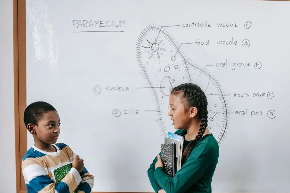
Unlock the profound secret behind growth, repair, and reproduction. Mitosis, the cellular process of division, orchestrates the precise duplication of genetic material, ensuring that every new cell is an exact replica of its parent. This intricate dance, vital for all eukaryotic life, is far more than just cell splitting; it is a meticulously choreographed sequence guaranteeing genetic fidelity across generations.
Dive deep with us as we dismantle the complexities of mitosis into clear, actionable insights. We will navigate each phase, from the initial preparations in interphase to the final separation, equipping you with the expert understanding typically reserved for seasoned biologists. We illuminate the crucial checkpoints, common pitfalls, and the astounding biological precision that underpins this fundamental process. To truly grasp this mechanism is to comprehend the fundamental units of life that compose all organisms. Prepare to forge a mastery of cellular division that transforms your appreciation for the very fabric of existence.
The Mitotic Imperative: Why Cells Divide with Precision
Mitosis stands as a foundational pillar of life, a dynamic process driving the growth, repair, and asexual reproduction of eukaryotic organisms. Every breath we take, every wound that heals, every new cell that replaces an old one—all are testaments to the flawless execution of mitosis. Our biological imperative dictates that cells must divide, not indiscriminately, but with absolute precision, ensuring that each daughter cell receives an identical set of chromosomes. This isn't merely about creating more cells; it's about maintaining genetic integrity, preventing errors that could lead to dysfunction, disease, or even organismal failure.
Imagine the immense challenge: a human cell contains approximately 3 billion base pairs of DNA, meticulously organized into 46 chromosomes. Duplicating this entire genome and then accurately distributing it into two new nuclei demands an unparalleled level of cellular orchestration. Without this exactitude, genetic information would be lost or unevenly distributed, compromising cellular function and potentially triggering uncontrolled growth, as seen in cancerous states. We forge our understanding by recognizing mitosis as a highly regulated, essential program, a testament to evolution's finest cellular engineering. This process is not a random event; it's a critical survival strategy for multicellularity and proliferation.
Interphase: The Crucial Prelude to Cellular Replication
Before the spectacular performance of mitosis can commence, the cell undergoes a period of intense preparation known as interphase. Often mistakenly viewed as a 'resting phase', interphase is anything but quiescent. It is a highly active and critical stage, comprising three distinct sub-phases: G1 (Gap 1), S (Synthesis), and G2 (Gap 2). During G1, the cell grows, synthesizes proteins, and carries out its normal metabolic functions. This is where the cell decides whether to proceed with division or enter a quiescent G0 state.
The S phase is the pivotal moment for genetic replication. Here, the cell meticulously duplicates its entire DNA content, ensuring that each chromosome now consists of two identical sister chromatids joined at a centromere. This DNA synthesis is error-checked and highly regulated, forming the bedrock of genetic fidelity for the subsequent division. Following DNA replication, the cell enters G2, a second growth phase where it synthesizes additional proteins and organelles, and further prepares for mitosis. Key checkpoints exist throughout interphase, acting as critical guardians that monitor DNA integrity and cellular readiness. We understand that a cell must be perfectly primed, its genetic material flawlessly duplicated, before it can even contemplate entering the intricate choreography of mitosis.
Prophase and Metaphase: Orchestrating Chromosome Alignment
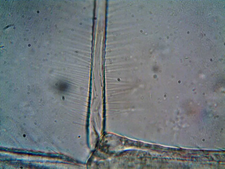
With interphase complete, the cell is poised for the drama of mitosis, which unfolds in a precise sequence of events. The first visible stage is Prophase. During prophase, the loosely packed chromatin fibers within the nucleus condense dramatically, becoming thousands of times more compact to form visible, discrete chromosomes. Each chromosome, now composed of two identical sister chromatids, becomes distinctly observable under a microscope. Simultaneously, the nuclear envelope, which encases the genetic material, begins to break down, and the nucleoli disappear. Crucially, the mitotic spindle, a complex structure of microtubules, starts to form, emanating from centrosomes that migrate to opposite poles of the cell.
Following prophase, the cell enters Metaphase, a phase renowned for its striking organization. The spindle microtubules, now fully formed, attach to the kinetochores – specialized protein structures located at the centromere of each sister chromatid. This attachment is not random; the microtubules actively pull and push the chromosomes until they meticulously align along the cell's equatorial plane, forming the 'metaphase plate.' This precise alignment is paramount. It ensures that when the sister chromatids finally separate, each daughter cell will receive an exact, full complement of genetic material. We observe metaphase as the ultimate test of the spindle's precision, a critical checkpoint ensuring equal distribution.
Anaphase and Telophase: The Separation and Rebuilding Phases
The meticulous alignment of chromosomes in metaphase sets the stage for the most dramatic event of mitosis: the separation of sister chromatids. This occurs during Anaphase. At the onset of anaphase, the cohesive proteins holding the sister chromatids together at the centromere are cleaved. This allows the sister chromatids to abruptly separate, instantly becoming individual chromosomes. These newly separated chromosomes are then rapidly pulled by the shortening spindle microtubules towards opposite poles of the cell. This movement is powered by motor proteins, consuming ATP to achieve precise and swift segregation. The cell visibly elongates during this phase, further facilitating the movement of chromosomes to their respective ends. Errors in anaphase, such as non-disjunction, lead to an uneven distribution of chromosomes, a common cause of genetic disorders.
As the chromosomes arrive at their designated poles, Telophase commences, effectively reversing many of the events of prophase. At each pole, a new nuclear envelope reforms around the now segregated sets of chromosomes. The chromosomes themselves begin to decondense, reverting to their less compact chromatin state. Concurrently, the mitotic spindle disassembles and disappears. Nucleoli reappear within the newly forming nuclei. By the end of telophase, the cell essentially contains two distinct, identical nuclei, each housing a complete and identical genetic blueprint. We recognize telophase as the completion of nuclear division, the meticulous rebuilding of two distinct genetic compartments within a single cell.
Cytokinesis: Finalizing the Split into Two Identical Cells
While telophase marks the completion of nuclear division (karyokinesis), the process of cell division is not truly finalized until the cytoplasm itself divides. This crucial step is known as Cytokinesis. Cytokinesis typically overlaps with the late stages of mitosis, often beginning during anaphase or telophase, and results in the formation of two physically separate, genetically identical daughter cells. The mechanism of cytokinesis differs significantly between animal cells and plant cells, reflecting their distinct structural organizations.
In animal cells, cytokinesis is initiated by the formation of a contractile ring composed of actin and myosin filaments. This ring forms just beneath the plasma membrane in the metaphase plate region. As the ring contracts, it pinches the cell inward, creating a 'cleavage furrow' that gradually deepens until the parent cell is completely divided into two. In contrast, plant cells, with their rigid cell walls, cannot form a cleavage furrow. Instead, vesicles originating from the Golgi apparatus migrate to the cell's equator, fusing to form a 'cell plate' that grows outward until it fuses with the existing plasma membrane and cell wall, effectively dividing the cell. We observe cytokinesis as the essential physical separation, concluding the magnificent journey of cell division and yielding two fully functional, independent biological units.
Mastering Mitosis: Common Pitfalls and Biological Precision
Our exploration reveals mitosis as a process of incredible precision, but what happens when this precision falters? Errors during mitosis can have profound consequences. A classic pitfall is non-disjunction, where sister chromatids fail to separate correctly during anaphase, leading to daughter cells with an abnormal number of chromosomes (aneuploidy). This is a primary cause of genetic disorders, such as Down syndrome (Trisomy 21). Another critical error arises from uncontrolled cell division, often due to failures in cell cycle checkpoints. This deregulation is the hallmark of cancer, where cells proliferate relentlessly, forming tumors and compromising organismal health.
To optimize our understanding and avoid these biological pitfalls, we must emphasize the role of checkpoints: molecular 'stop signs' that ensure all conditions are met before proceeding to the next phase. The G1, G2, and M-phase (spindle assembly) checkpoints are vital guardians of genetic integrity. Insider tip: Visualizing each phase with key identifiers (e.g., condensed chromosomes at the equator for metaphase, separating chromatids for anaphase) is crucial for rapid identification. We forge a comprehensive view of mitosis not just by understanding its ideal progression, but also by recognizing the robust mechanisms in place to correct or halt faulty division, underscoring the vital balance between growth and control in cellular biology.
Key Takeaways
Mitosis Fundamentals
Mitosis is the process of nuclear division in eukaryotic cells, resulting in two genetically identical daughter cells. It is essential for growth, repair, and asexual reproduction, ensuring genetic fidelity. The overall goal is to precisely distribute duplicated chromosomes equally between new cells.
Interphase: The Preparation
Before mitosis, cells undergo Interphase (G1, S, G2). In S phase, DNA replicates, forming two sister chromatids per chromosome. G1 and G2 involve cell growth and synthesis of necessary proteins and organelles. Interphase is crucial for preparing the cell for accurate division.
Mitotic Phases (PMAT)
- Prophase: Chromosomes condense, nuclear envelope breaks down, mitotic spindle forms.
- Metaphase: Chromosomes align at the metaphase plate, ensuring equal distribution.
- Anaphase: Sister chromatids separate, becoming individual chromosomes, and move to opposite poles.
- Telophase: New nuclear envelopes form around chromosome sets, chromosomes decondense, spindle disappears.
Cytokinesis: Cytoplasmic Division
- Animal Cells: A 'cleavage furrow' forms via a contractile ring, pinching the cell in two.
- Plant Cells: A 'cell plate' forms from fusing vesicles, growing outwards to divide the cell and build a new cell wall.
This process physically separates the two new nuclei into distinct daughter cells.
Importance and Precision
Mitosis relies on strict cell cycle checkpoints to prevent errors. Failures, such as non-disjunction, can lead to aneuploidy (e.g., Down syndrome) or uncontrolled cell proliferation (cancer). The precision of mitosis is vital for maintaining organismal health and genetic stability.
FAQ
-
What is the primary goal of mitosis?
The primary goal of mitosis is to produce two daughter cells that are genetically identical to the parent cell. This ensures that each new cell receives a complete and exact set of chromosomes, maintaining genetic fidelity essential for growth, tissue repair, and asexual reproduction.
-
How does cytokinesis differ in animal and plant cells?
In animal cells, cytokinesis occurs via the formation of a 'cleavage furrow,' a contractile ring of actin and myosin filaments that pinches the cell in two. In plant cells, which have rigid cell walls, a 'cell plate' forms in the middle of the cell from fusing vesicles, growing outward to divide the cytoplasm and eventually forming a new cell wall.
-
Why is interphase considered a critical stage despite not being part of mitosis?
Interphase is critical because it is the period of intense cell growth, organelle synthesis, and most importantly, DNA replication (S phase). Without the accurate duplication of genetic material during interphase, mitosis would result in genetically incomplete or incorrect daughter cells, compromising cell function and potentially leading to disease.
-
What are the consequences if mitosis goes wrong?
Errors in mitosis can have severe consequences. Common errors like non-disjunction (unequal chromosome distribution) lead to aneuploidy, causing genetic disorders. Uncontrolled or faulty mitosis, often due to failures in cell cycle checkpoints, is a fundamental characteristic of cancer, leading to abnormal cell proliferation and tumor formation.