
> Basic Biology Concepts > Cell Structure and Cellular Organization > Dissect Structural Differences in Bacterial and Eukaryotic Cells
Dissect Structural Differences in Bacterial and Eukaryotic Cells
We forge our understanding of life by exploring the fundamental structures that drive all forms of life. At the heart of this quest lies an essential duality: the distinction between bacterial (prokaryotic) cells and eukaryotic cells. This architectural divergence, forged over billions of years of evolution, is not merely a biological curiosity; it is the key to understanding cells as the fundamental units of life and unlocking vital strategies in medicine, biotechnology, and ecology. Indeed, the ability to differentiate these two cellular architectures revolutionizes our approach to infectious diseases, the development of new antibiotics, and even genetic modification.
In this article, we initiate a surgical exploration of the key structural disparities that separate these two cellular categories. We go beyond surface-level definitions to delve into the intimate mechanisms, specialized components, and functional implications of each distinction. Prepare to optimize your perspective on cell biology and forge undeniable expertise. Understanding these differences is not just about learning facts; it is about acquiring a powerful lens to interpret the biological world around us with expert precision.
Unveiling the Fundamental Divide: Nucleus and Domains of Life
At the very core of cellular biology, the distinction between prokaryotic and eukaryotic cells represents one of life's most profound evolutionary splits. The defining characteristic, a concept we must engrave into our foundational knowledge, is the presence or absence of a true nucleus. Eukaryotic cells, a term derived from Greek meaning 'true nucleus,' house their genetic material within a sophisticated, membrane-bound compartment. This nuclear envelope, perforated by nuclear pores, meticulously regulates the passage of molecules, ensuring precise control over gene expression and DNA replication. This compartmentalization is not merely an aesthetic choice; it dramatically elevates the complexity of cellular regulation and function.
Conversely, prokaryotic cells, meaning 'before nucleus,' lack this central organizing principle. Their genetic material—typically a single, circular chromosome—resides in a region of the cytoplasm known as the nucleoid. This lack of a membrane barrier implies a much more direct coupling of transcription and translation, allowing for incredibly rapid responses to environmental changes. This fundamental difference in genetic organization dictates vastly different strategies for genetic management, repair, and expression, impacting everything from growth rates to adaptability.
We observe this distinction across the entire tree of life: Bacteria and Archaea constitute the prokaryotic domains, while Eukarya encompasses animals, plants, fungi, and protists. This foundational divergence impacts cellular size, with eukaryotic cells typically being 10 to 100 times larger in volume than their prokaryotic counterparts. This size disparity is a direct consequence of internal organization. The eukaryotic advantage lies in its ability to manage a larger internal volume through internal compartmentalization, allowing for a division of labor that prokaryotes achieve through sheer numbers and rapid reproduction. Understanding this initial divide is our first tactical step in mastering cellular architecture.
Deciphering Internal Complexity: Organelles and Cytoskeletal Architecture
Beyond the nucleus, the internal landscape of eukaryotic cells is a veritable city of specialized structures, each performing dedicated functions—these are the membrane-bound organelles. We encounter the mitochondria, the powerhouses of the cell, orchestrating cellular respiration; the endoplasmic reticulum (ER), a vast network involved in protein synthesis and lipid metabolism; and the Golgi apparatus, responsible for modifying, sorting, and packaging proteins and lipids for secretion or delivery to other organelles. Lysosomes manage cellular waste, while peroxisomes handle detoxification. This intricate division of labor within eukaryotes optimizes biochemical processes by concentrating specific enzymes and substrates, minimizing interference, and maximizing efficiency across a larger cellular volume.
Prokaryotic cells, in stark contrast, conspicuously lack these membrane-bound organelles. Their metabolic activities, including respiration and photosynthesis (in some species), occur on the plasma membrane itself or within the cytoplasm. While they possess ribosomes for protein synthesis, these are structurally distinct (70S) from the larger eukaryotic ribosomes (80S), a critical difference often exploited in antibiotic design. The absence of internal compartments in prokaryotes demands alternative strategies for efficiency, often relying on rapid diffusion, high surface-area-to-volume ratios, and the localized concentration of enzymes within specialized regions of the cytoplasm or associated with the cell membrane.
Another crucial differentiator is the cytoskeleton. Eukaryotic cells possess a highly dynamic and complex cytoskeleton composed of microtubules, microfilaments, and intermediate filaments. This network is instrumental in maintaining cell shape, enabling cell movement, facilitating intracellular transport of vesicles and organelles, and orchestrating cell division (mitosis and meiosis). While prokaryotes were historically thought to lack a cytoskeleton, we now know they possess rudimentary, homologous protein structures (e.g., FtsZ, MreB) that play vital roles in cell division, shape maintenance, and chromosome segregation. However, their complexity and functional versatility remain significantly less evolved than their eukaryotic counterparts. We must recognize this difference as a pivotal factor in the distinct mobility and structural integrity observed across the two cell types.
Charting Genetic Landscapes: Chromosomes, Plasmids, and Replication Mechanisms
The manner in which genetic information is stored, organized, and duplicated represents another profound divergence. In eukaryotic cells, the genetic blueprint is meticulously packaged into multiple linear chromosomes, each composed of DNA tightly coiled around histone proteins. These histones play a vital role in chromatin condensation, gene regulation, and DNA repair. The entire set of chromosomes is confined within the nucleus, and its duplication and segregation during cell division are orchestrated by the complex processes of mitosis and meiosis, ensuring precise distribution to daughter cells.
Prokaryotic cells, on the other hand, typically possess a single, circular chromosome located in the nucleoid region. This chromosome is generally not associated with histones in the same manner as eukaryotes, though histone-like proteins do assist in DNA compaction. A hallmark of prokaryotic genetics is the frequent presence of plasmids—small, independent, circular DNA molecules that can carry advantageous genes, such as antibiotic resistance or virulence factors. These plasmids can be exchanged between bacteria through horizontal gene transfer, providing a rapid mechanism for adaptation and evolution, a strategy largely absent in the same form in eukaryotes.
The replication strategies also diverge significantly. Prokaryotic cells undergo binary fission, a relatively simple and rapid process where the single chromosome duplicates, and the cell divides into two identical daughter cells. This speed enables bacterial populations to expand exponentially under favorable conditions. Eukaryotic cells, with their multiple linear chromosomes and nuclear envelope, require the elaborate machinery of mitosis (for somatic cells) or meiosis (for germ cells) to ensure accurate segregation. These processes involve complex spindle fibers, checkpoints, and nuclear envelope breakdown and reformation, highlighting the increased investment in genetic fidelity required for larger, multicellular organisms. Understanding these genetic architectural blueprints is crucial for comprehending their respective evolutionary trajectories and adaptive capacities.
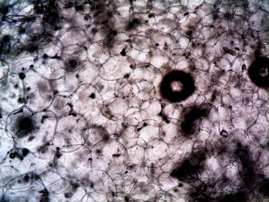
Beyond the Boundary: Cell Walls, Membranes, and External Appendages
The outermost layers of cells—the boundaries that interact with the external environment—also exhibit striking differences. All cells possess a plasma membrane, a universal lipid bilayer that regulates the passage of substances. However, eukaryotic membranes often contain sterols (like cholesterol in animal cells), which modulate fluidity and stability. Prokaryotic membranes typically lack sterols, though some may contain structurally analogous molecules called hopanoids. These lipid compositional differences influence membrane fluidity, permeability, and resistance to environmental stress, reflecting adaptations to diverse habitats.
A more prominent difference lies in the cell wall. Most prokaryotic cells, particularly bacteria, possess a rigid cell wall primarily composed of peptidoglycan (murein). This unique polymer, a sugar-amino acid meshwork, provides structural support, protects against osmotic lysis, and is the target of many antibiotics (e.g., penicillin). The thickness and composition of peptidoglycan vary between Gram-positive and Gram-negative bacteria, a critical distinction for bacterial identification and treatment. Eukaryotic cell walls are present in plants (cellulose), fungi (chitin), and some protists, but are completely absent in animal cells. These eukaryotic cell walls differ fundamentally in chemical composition and structural organization from their prokaryotic counterparts.
Finally, we examine external appendages. Many prokaryotes utilize structures like flagella for motility, propelling the cell through liquid environments. Bacterial flagella are composed of the protein flagellin and rotate like propellers, powered by a proton motive force. This is fundamentally different from eukaryotic flagella and cilia, which are composed of microtubules and exhibit a whip-like motion, powered by ATP. Prokaryotes also feature pili (fimbriae), short, hair-like appendages crucial for adhesion to surfaces and host cells, and specialized sex pili for bacterial conjugation (transfer of genetic material). Some bacteria may also possess a protective capsule—a thick, sticky polysaccharide layer external to the cell wall—that enhances virulence and provides protection against phagocytosis. These diverse external structures equip bacteria for survival and interaction within their specific ecological niches, offering compelling targets for therapeutic intervention and ecological studies.
Key Takeaways
Fundamental Distinction: Nucleus
Eukaryotic cells possess a membrane-bound nucleus containing linear chromosomes. Prokaryotic cells lack a nucleus; their circular chromosome is in the nucleoid.
Internal Organization: Organelles
Eukaryotes feature extensive membrane-bound organelles (mitochondria, ER, Golgi) for compartmentalized functions. Prokaryotes lack these, with metabolic activities often occurring on the plasma membrane.
Genetic Material & Replication
Eukaryotes have multiple linear chromosomes with histones, undergoing mitosis/meiosis. Prokaryotes typically have a single circular chromosome and often plasmids, replicating via binary fission.
Cell Walls & External Features
Bacterial cell walls are primarily composed of peptidoglycan. Eukaryotic cell walls (if present) are chemically distinct (cellulose, chitin) or absent (animals). Prokaryotes often have specialized flagella, pili, and capsules not found in eukaryotes.
Ribosomes & Cytoskeleton
Both have ribosomes, but prokaryotic ribosomes are 70S, while eukaryotic are 80S. Eukaryotes possess a complex cytoskeleton; prokaryotes have simpler, homologous protein structures.
FAQ
-
What is the single most defining structural difference between prokaryotic and eukaryotic cells?
The single most defining structural difference is the presence of a membrane-bound nucleus in eukaryotic cells, which encloses their genetic material. Prokaryotic cells lack a true nucleus, with their genetic material located in a region called the nucleoid.
-
Do bacterial cells possess ribosomes, and are they similar to those in eukaryotic cells?
Yes, bacterial cells do possess ribosomes, which are essential for protein synthesis. However, prokaryotic ribosomes are 70S, while eukaryotic ribosomes are larger at 80S. This size difference is often exploited by antibiotics to selectively target bacterial protein synthesis without harming host cells.
-
Why is understanding these structural differences crucial for medical science?
Understanding these differences is paramount for medical science because many antibacterial drugs (antibiotics) selectively target structures unique to bacteria, such as their peptidoglycan cell wall (e.g., penicillin) or their 70S ribosomes (e.g., erythromycin). This specificity allows for effective treatment of bacterial infections with minimal harm to human (eukaryotic) cells. It also informs vaccine development and understanding pathogenesis.
-
Are plasmids unique to prokaryotic cells, and what is their significance?
While some eukaryotic organisms can have plasmid-like DNA, large, self-replicating plasmids are overwhelmingly characteristic of prokaryotic cells. Their significance is immense: they often carry genes that confer advantageous traits, such as antibiotic resistance, heavy metal tolerance, or virulence factors, and can be readily exchanged between bacteria via horizontal gene transfer, contributing significantly to bacterial adaptation and evolution.