
> Laboratory Techniques > PCR and DNA Amplification Methods > Maîtriser les Distinctions : PCR et qPCR pour la Biologie Moléculaire
Maîtriser les Distinctions : PCR et qPCR pour la Biologie Moléculaire
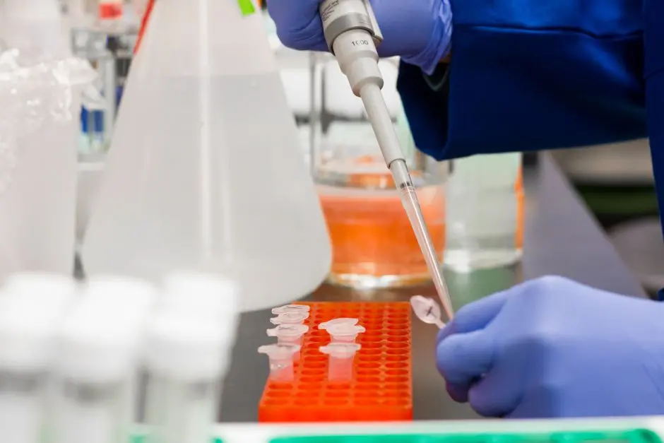
In the field of molecular biology, the ability to manipulate and analyze DNA is fundamental. Among the most revolutionary tools, Polymerase Chain Reaction (PCR) and quantitative PCR (qPCR) stand out for their power and versatility. These techniques, while sharing a common amplification principle, unlock distinct levels of information, ranging from simple detection to precise quantification of genetic sequences. Understanding their fundamental differences is not merely an academic requirement; it is a strategic skill that dictates the direction of research projects, clinical diagnostics, and biotechnological applications.
This article undertakes a surgical exploration of the nuances between PCR and qPCR. We dissect their mechanisms, optimal applications, and pitfalls to avoid, thereby equipping practitioners with the necessary knowledge to select the most suitable tool for their objectives. Prepare to delve into the heart of DNA amplification, optimize your protocols, and decipher data with unparalleled accuracy. We reveal how these methods integrate into the broader ecosystem of modern PCR techniques for DNA amplification and analysis, transforming our approach to biology and health.
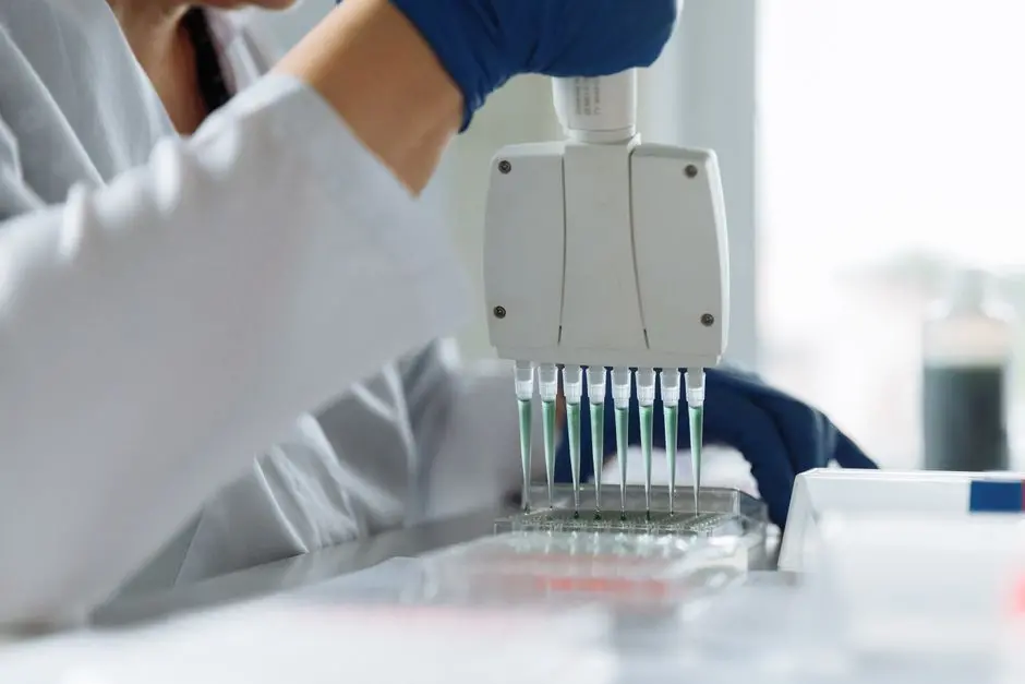
Demystifying Standard PCR: Fundamentals of Qualitative Amplification
Standard Polymerase Chain Reaction (PCR) is the cornerstone of DNA amplification techniques, an innovation that has reshaped molecular biology. Our objective is clear: to generate millions of copies of a specific DNA sequence from even a minimal sample. This process revolves around a repetitive and precisely controlled thermal cycle, leveraging the thermostability of a DNA polymerase enzyme, such as Taq polymerase.
The PCR cycle breaks down into three critical steps:
- Denaturation (94-98°C): We heat the DNA sample to separate the two strands of the double helix, making targets accessible.
- Annealing (50-65°C): We lower the temperature, allowing specific primers (short synthetic DNA fragments) to bind to their complementary sequences on the single-stranded DNA strands. The annealing temperature is a critical parameter that we optimize to ensure specificity.
- Extension (72°C): At this optimal temperature, Taq polymerase comes into play, synthesizing a new DNA strand using the primer as a starting point and the original DNA strand as a template.
This cycle is typically repeated 25 to 40 times, leading to exponential amplification of the target sequence. The final product, an amplicon, is usually visualized by agarose gel electrophoresis, where the presence of a band of expected size confirms amplification. Standard PCR is primarily a qualitative or semi-quantitative method, indicating the presence or absence of a sequence, or allowing a rough estimation of its relative amount by band intensity. We utilize it for DNA cloning, genotyping, pathogen detection, and sample preparation for next-generation sequencing. Mastery of primer, DNA, dNTPs, and MgCl₂ concentrations is imperative to minimize artifacts such as primer dimers and non-specific amplifications, which compromise the validity of our results.
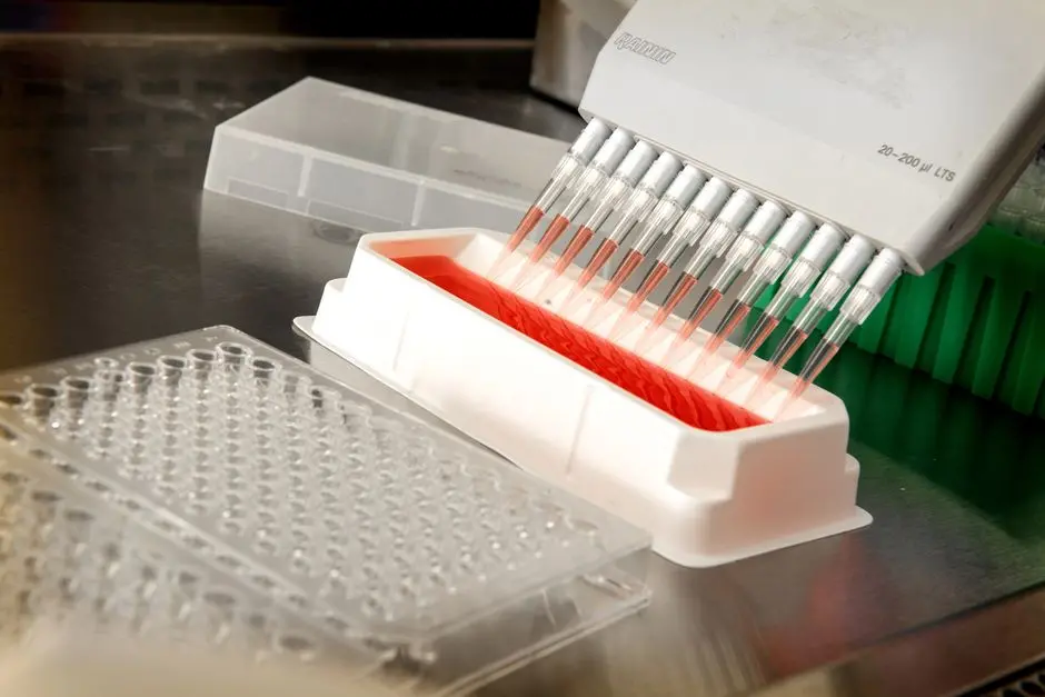
Deploying qPCR: The Power of Real-Time Quantification
Quantitative PCR, or qPCR, transcends standard PCR by introducing the ability to measure DNA amplification in real-time. This major advancement allows us not only to detect a sequence but also to quantify it with exceptional precision. The fundamental principle of qPCR integrates fluorescence detection, providing us with a dynamic readout of amplicon accumulation in each cycle.
We employ two primary strategies for fluorescent detection in qPCR:
- DNA-binding Dyes (e.g., SYBR Green): These dyes specifically bind to double-stranded DNA, emitting fluorescence only in this form. As the target DNA amplifies, the amount of double-stranded DNA increases, leading to a proportional rise in the fluorescent signal. The advantage lies in their cost-effectiveness and simplicity, as they do not require specific probes. However, they can bind to any double-stranded DNA, including non-specific products, necessitating a post-PCR melt curve analysis to verify specificity.
- Sequence-Specific Probes (e.g., TaqMan, Molecular Beacons): These probes are oligonucleotides labeled with a fluorophore and a quencher. As long as the probe is intact, the quencher suppresses the fluorophore's signal. During the elongation phase, Taq polymerase, with its 5' > 3' exonuclease activity, degrades the bound probe, releasing the fluorophore from the quencher and allowing fluorescence emission. This method offers superior specificity because fluorescence is generated only upon degradation of the target-specific probe.
The crucial point in qPCR is the cycle threshold (Ct), also known as the quantification cycle (Cq). It represents the number of cycles required for the sample's fluorescent signal to exceed the background noise. A lower Ct value indicates a higher initial quantity of the target sequence. We leverage the inverse relationship between Ct and initial target DNA concentration to perform absolute (via a standard curve) or relative quantifications (by comparison with a reference gene). qPCR is indispensable for gene expression analysis (after reverse transcribing RNA to cDNA), viral load quantification, mutation detection, and high-throughput screening. Our rigor in primer and probe design, along with strict sample quality control, is paramount to ensuring the reliability of quantitative data.
Discerning Architectures: Key Differences Between PCR and qPCR
While PCR and qPCR share the objective of amplifying DNA sequences, their technological architectures and analytical capabilities diverge radically. Understanding these distinctions is crucial for orchestrating biological experiments with maximum efficiency and flawless data interpretation. We will dissect the major contrasts that define the utility of each method.
- Timing of Measurement: The most fundamental difference lies in the timing of detection. Standard PCR is an "endpoint" technique: we analyze the amplicon only after all amplification cycles are completed. This gives us a static snapshot of the final product. In contrast, qPCR is a "real-time" technique: we monitor amplicon accumulation with each cycle, as the reaction progresses. This dynamic approach allows us to capture the exponential phase of amplification, where the correlation between fluorescence and DNA quantity is most linear and most representative of the initial amount.
- Nature of Information Provided: Standard PCR provides qualitative (presence/absence of a sequence) or semi-quantitative (rough estimation based on band intensity) information. It excels at visually confirming amplification. qPCR, on the other hand, delivers precise quantitative data, enabling us to determine the initial copy number of a target sequence with high sensitivity and a wide dynamic range. We obtain not just a "yes/no" answer, but also a "how much."
- Equipment Cost and Complexity: A standard thermocycler is the sole equipment required for PCR. Post-reaction analysis necessitates a gel electrophoresis system. qPCR, conversely, demands a specialized thermocycler equipped with an optical detection system capable of measuring fluorescence in real time. This equipment is generally more expensive, and data management requires dedicated analysis software. Reagents for qPCR (probes, optimized master mixes) are also often more costly than those for standard PCR.
- Sensitivity and Specificity: qPCR is intrinsically more sensitive than standard PCR, capable of detecting lower amounts of DNA. Real-time detection also reduces the risk of post-PCR contamination, as tubes remain sealed after reagent addition. With the use of specific probes, qPCR also offers improved specificity compared to interfering dyes or simple size analysis in PCR.
In summary, PCR reveals what is present, while qPCR reveals how much and with what kinetics. Our choice between these two techniques is a strategic decision, determined by the precise biological questions we seek to answer and the resources at our disposal.
Application Strategies: When to Choose PCR or qPCR for Experimental Excellence
The choice between standard PCR and qPCR is not arbitrary; it is the result of a rigorous analysis of experimental objectives and inherent constraints. We forge this decision by evaluating the fundamental biological question we are asking. Each technique is a powerful tool, but its true value shines when applied to its intended domain, thereby optimizing our resources and accelerating our discoveries.
- When to Deploy Standard PCR: We favor standard PCR when our primary objective is the qualitative detection or simple amplification of a DNA sequence. For example, for gene cloning, where we need to generate large quantities of a specific DNA fragment for subsequent manipulation (ligation, insertion into vectors). In routine diagnostics, PCR is sufficient to confirm the presence of a pathogen if quantification is not critical. Genotyping, polymorphism detection by RFLP, or marker amplification for DNA fingerprinting are other ideal applications. Its ease of implementation, lower cost, and robustness make it the method of choice for quick checks and sample preparations. However, we must remain vigilant about yield variability and the risk of false negatives or positives if the protocol is not meticulously optimized.
- When to Engage qPCR: qPCR becomes the indispensable tool when precise quantification and sensitivity are the critical imperatives of our research. For gene expression analysis, where we need to measure changes in mRNA levels under different physiological or pathological conditions, qPCR (after reverse transcription into cDNA, RT-qPCR) is unparalleled. In clinical virology, quantification of viral load (e.g., HIV, hepatitis B/C) is vital for monitoring disease progression and treatment efficacy. The detection of low levels of DNA or RNA, for instance, in liquid biopsies for cancer screening or rare cell detection, capitalizes on qPCR's superior sensitivity. We also use qPCR for transgene detection, DNA chip validation, and copy number variation (CNV) analysis. Its high-throughput potential and automated data analysis reinforce its role in high-volume clinical and research laboratories.
For both techniques, rigorous primer design is non-negotiable. For qPCR, establishing reliable standard curves, judicious selection of reference genes (for relative quantification), and validation of amplification efficiency are crucial steps. We invest in the continuous training of our teams in best practices and the latest advancements to maximize the relevance and robustness of our data.
Key Takeaways
PCR Standard : Détection Qualitative
Standard PCR amplifies target DNA sequences through cycles of denaturation, annealing, and extension. It is used for qualitative (presence/absence) or semi-quantitative detection of a sequence. Its analysis is performed at 'end-point', typically by gel electrophoresis. Ideal for cloning, genotyping, and simple pathogen detection. More economical and simpler to implement.
qPCR: Precise Real-Time Quantification
qPCR (quantitative PCR) allows for real-time DNA quantification through fluorescence detection generated during amplification. The fluorescent signal is proportional to the amplicon quantity, and the threshold cycle (Ct) is inversely proportional to the initial amount of target DNA. It is crucial for gene expression analysis, viral load quantification, and mutation detection. It offers increased sensitivity and specificity but requires specialized equipment and more expensive reagents.
Key Differences and Choice Strategy
PCR is a qualitative/semi-quantitative 'endpoint' measurement, whereas qPCR is a quantitative 'real-time' measurement. qPCR is more sensitive, specific, and offers a wider dynamic range. The choice between the two depends on experimental goals: PCR for simple detection or bulk amplification; qPCR for precise quantification of expression or DNA/RNA quantity. Rigor in protocol design is vital for both methods.
FAQ
-
What is the main advantage of qPCR over standard PCR?
The fundamental advantage of qPCR lies in its ability to precisely quantify the initial amount of DNA (or RNA after reverse transcription) present in a sample. Unlike standard PCR which provides qualitative endpoint detection, qPCR monitors amplification in real-time, allowing for the determination of initial copy numbers with high sensitivity and a wide dynamic range. -
Can standard PCR be used for gene expression quantification?
Standard PCR is not suitable for precise gene expression quantification. It is qualitative or, at best, semi-quantitative (based on band intensity on a gel), which does not allow for reliable discrimination of small expression differences. For reliable gene expression quantification, qPCR (RT-qPCR) is the method of choice, as it measures cDNA accumulation in real-time and allows for precise numerical data to be obtained. -
What is the cycle threshold (Ct) in qPCR and why is it important?
The threshold cycle (Ct), or quantification cycle (Cq), is the number of amplification cycles at which the fluorescent signal of a sample exceeds a defined detection threshold, which is above background noise. Its value is inversely proportional to the initial amount of target DNA: a low Ct indicates a large amount of starting DNA, and vice versa. The Ct is the key parameter used for quantification in qPCR. -
What are the different types of fluorescent detection used in qPCR?
We mainly use two types of fluorescent detection methods in qPCR: DNA intercalating dyes (such as SYBR Green), which bind non-specifically to all double-stranded DNA and emit fluorescence, and specific probes (such as TaqMan probes), which are oligonucleotides labeled with a fluorophore and a quencher, and whose fluorescence is only released after their specific degradation by polymerase.
-
Is qPCR more expensive than standard PCR?
Yes, qPCR is generally more expensive than standard PCR. It requires specialized equipment (thermocycler with an optical detection system), and the reagents (specific probes, optimized master mixes) are often more costly. However, the additional cost is justified by the richness of quantitative information and increased sensitivity it offers, which can prevent additional experiments and optimize laboratory time.