
> Laboratory Techniques > Microscopy and Cellular Imaging > Amplify Biological Insight: Confocal's Edge Over Traditional Microscopy
Amplify Biological Insight: Confocal's Edge Over Traditional Microscopy
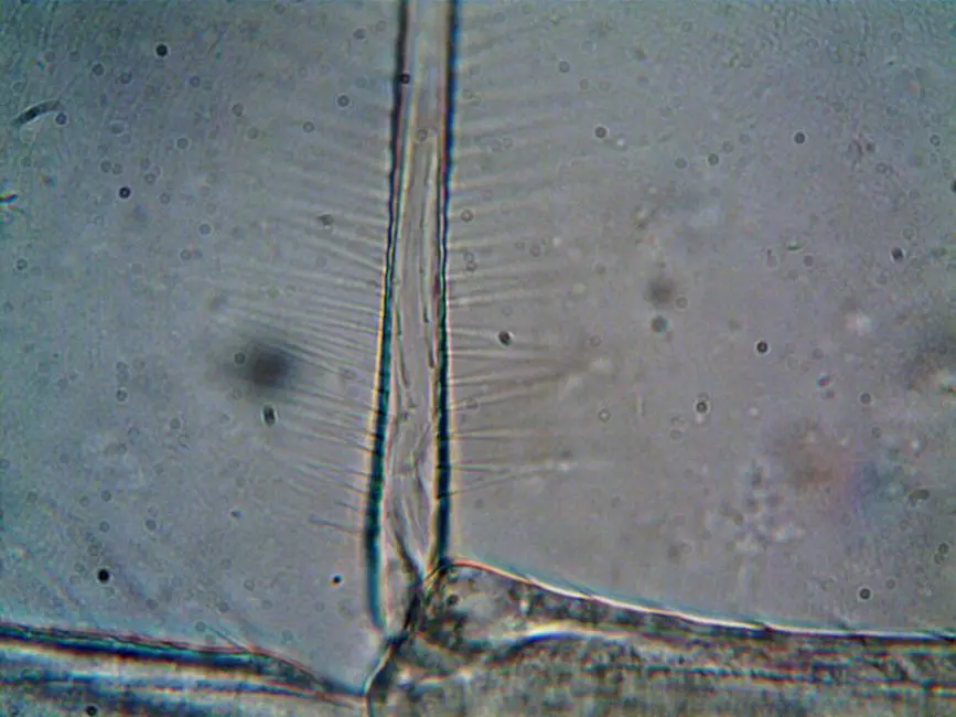
The evolution of microscopy has dramatically reshaped our comprehension of the biological world. While traditional methods offered foundational insights, the advent of techniques like confocal microscopy propels us into an era of unprecedented clarity. This article delves deep into the transformative advantages that confocal microscopy delivers over its predecessors, fundamentally redefining how we visualize cellular structures and dynamic processes. We will dissect the optical principles, explore advanced applications, and equip you with the knowledge to harness its full potential, ensuring your research benefits from the sharpest possible views.
For a broader understanding of advanced laboratory imaging methods for biological structures, this exploration is indispensable for researchers seeking to push the boundaries of biological discovery. Prepare to optimize your experimental design and unlock biological opportunities previously obscured by conventional limitations.
Deciphering the Constraints of Traditional Microscopy
Traditional microscopy, encompassing techniques such as brightfield, phase contrast, and widefield fluorescence, has undeniably formed the bedrock of cellular biology for centuries. These methods have enabled monumental discoveries, from the identification of cells to the observation of fundamental biological processes. However, their inherent optical limitations often present significant barriers to advanced research, particularly when investigating complex, three-dimensional biological samples. We frequently encounter issues that compromise image quality and interpretability, impeding precise scientific inquiry.
A primary challenge with widefield fluorescence microscopy, for instance, involves the capture of out-of-focus light. When illuminating a thick specimen, light from above and below the focal plane simultaneously reaches the detector, creating a pervasive haze. This 'blur' significantly reduces image contrast and resolution, making it nearly impossible to distinguish fine cellular structures or quantify fluorescent signals accurately. Furthermore, the extensive illumination required for widefield imaging can lead to increased phototoxicity and photobleaching, especially during prolonged observation or live-cell imaging experiments. Phototoxicity damages living cells, altering their physiological state, while photobleaching irreversibly destroys fluorescent markers, shortening observation windows. These factors collectively constrain our ability to perform high-fidelity, long-term studies, thereby limiting the depth of biological insight we can achieve. Traditional methods, while foundational, compel us to seek sharper, more controlled imaging solutions.
The Confocal Revolution: Precision Through Optical Sectioning
Confocal microscopy fundamentally transforms biological imaging by introducing the concept of optical sectioning, a revolutionary mechanism that precisely rejects out-of-focus light. This ingenious approach relies on a meticulously positioned pinhole aperture in the detection pathway, strategically placed in a conjugate plane (confocal) to the specimen focal point. Only light originating from the focal plane can pass through this pinhole and reach the detector; light from out-of-focus planes is blocked. This optical filtering dramatically enhances image clarity and contrast, delivering crisp, high-resolution images previously unattainable with widefield techniques.
We gain unparalleled control over the depth of focus, allowing us to acquire images from discrete optical slices within a thick specimen. This capability is pivotal for generating stunning three-dimensional reconstructions of cells, tissues, and even entire small organisms. The axial resolution, or resolution along the Z-axis, is substantially improved, typically achieving values in the range of 500-800 nanometers, significantly better than the several micrometers characteristic of widefield methods. This precision reveals intricate cellular architectures and subcellular localization with unprecedented detail. The point-scanning nature of confocal microscopy, where a focused laser beam scans across the sample pixel by pixel, also contributes to a superior signal-to-noise ratio, as each point is illuminated and detected individually. This targeted approach minimizes background noise and maximizes the detection of true fluorescent signals, ensuring robust and quantifiable data collection for every experiment we undertake.
Unlocking Advanced Capabilities: Dynamics, Dimensions, and Diversity
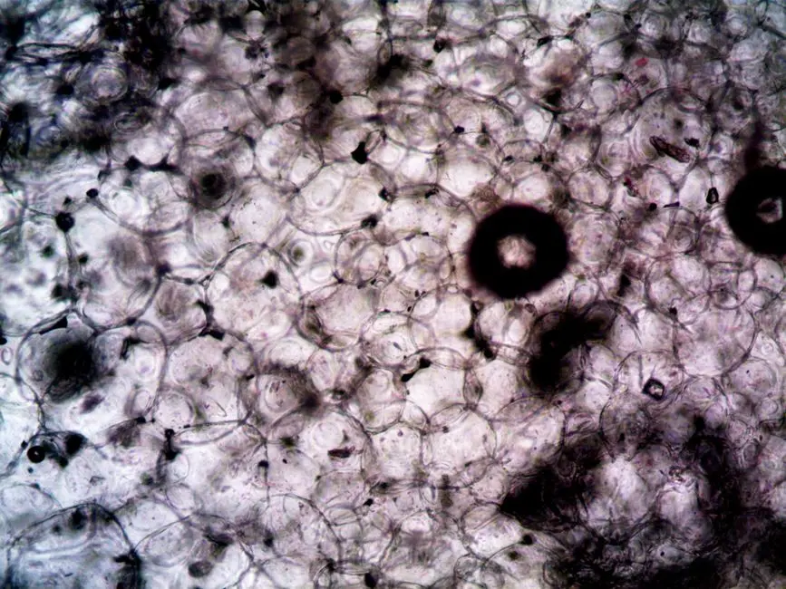
Confocal microscopy extends beyond mere image clarity, unlocking a suite of advanced capabilities crucial for cutting-edge biological research. Its ability to generate precise optical sections empowers us to construct intricate three-dimensional models of biological structures with exceptional fidelity. We can stack these 2D slices to visualize organelles, cellular networks, and tissue architectures in their true spatial context, fostering a deeper understanding of their relationships and functions. This volumetric data is indispensable for fields like developmental biology, neuroscience, and tissue engineering, where spatial organization dictates biological outcomes.
Furthermore, confocal microscopy excels in live-cell imaging, allowing us to observe dynamic cellular processes in real-time. While considerations for phototoxicity remain, modern confocal systems incorporate features like reduced laser power, optimized scanning strategies, and sensitive detectors to minimize cellular stress. This enables longitudinal studies of processes such as protein trafficking, cellular migration, and receptor dynamics within living systems. We also leverage confocal's power for multi-labeling experiments. By using multiple lasers and spectrally distinct fluorophores, we can simultaneously image several targets within a single cell or tissue. The pinhole's ability to reject crosstalk between channels ensures accurate co-localization studies, providing insights into complex molecular interactions. Our expertise lies in selecting optimal fluorophore combinations and mastering acquisition parameters to prevent photobleaching and maximize signal integrity, thereby capturing the full spectrum of cellular activity.
Optimizing Confocal Potential: Best Practices and Pitfalls to Avoid
While confocal microscopy offers unparalleled advantages, mastering its potential demands strategic planning and a keen awareness of best practices. Initially, we recognize the investment in confocal systems can be substantial, both in terms of cost and the learning curve. However, the return on this investment in data quality and depth of insight is immense. Our primary focus must center on meticulous sample preparation, ensuring optimal labeling and mounting to minimize aberrations and scattering. The choice of objective lens is critical; we prioritize high numerical aperture (NA) objectives for superior resolution and brightness, often balancing this with appropriate working distances for our specific samples.
For image acquisition, we meticulously optimize parameters. Setting appropriate laser power and detector gain is crucial to avoid oversaturation—a common error that obliterates quantitative data—or underexposure, which yields noisy images. We precisely adjust pixel size (zoom factor) to meet Nyquist sampling criteria, preventing aliasing and ensuring true resolution capture. Managing photobleaching, especially in live-cell imaging, involves minimizing laser exposure time, employing less photostable fluorophores, and using anti-fade reagents where appropriate. We also acknowledge that confocal scanning can be slower than widefield, demanding thoughtful experimental design for high-throughput applications. Understanding these nuances, and iteratively refining our protocols, allows us to mitigate common errors—such as incorrect pinhole settings, which compromise optical sectioning, or improper fluorophore selection leading to spectral bleed-through—thereby fully harnessing the confocal microscope's formidable power for robust and reliable biological discovery.
Key Takeaways
Elimination of Out-of-Focus Blur
Confocal microscopy employs a pinhole to block light from above and below the focal plane, eliminating blur prevalent in widefield microscopy. This delivers significantly clearer, higher-contrast images of biological samples.
Enhanced Axial Resolution and 3D Reconstruction
The ability to optically section specimens provides superior axial (Z-axis) resolution (500-800 nm). This is crucial for creating precise and detailed three-dimensional reconstructions of cellular and tissue structures, revealing complex spatial relationships.
Advanced Capabilities: Live-Cell Imaging and Multi-labeling
Confocal systems enable high-resolution live-cell imaging of dynamic processes and robust multi-labeling experiments without significant spectral crosstalk. This allows simultaneous visualization of multiple cellular components, critical for understanding complex biological interactions.
Optimizing Performance and Avoiding Pitfalls
Maximizing confocal benefits requires meticulous sample preparation, judicious objective selection, and precise parameter optimization (laser power, gain, pixel size). Careful management of phototoxicity and photobleaching is essential for quantitative, artifact-free data.
FAQ
-
What is the primary advantage of confocal microscopy over traditional widefield fluorescence?
The primary advantage of confocal microscopy lies in its ability to perform optical sectioning. This means it effectively rejects out-of-focus light, producing sharp, high-contrast images from specific focal planes within a thick sample. Traditional widefield microscopy, in contrast, captures light from all depths, resulting in blurry images with reduced resolution due to out-of-focus haze.
-
How does confocal microscopy achieve optical sectioning?
Confocal microscopy achieves optical sectioning through the strategic placement of a pinhole aperture in the detection pathway. This pinhole is positioned confocally (in a conjugate plane) with the illuminated focal point in the specimen. Only light originating from this precise focal plane can pass through the pinhole and reach the detector, while light from above or below the focal plane is blocked, effectively eliminating blur.
-
Is confocal microscopy suitable for live-cell imaging, and what are the considerations?
Yes, confocal microscopy is highly suitable for live-cell imaging, offering excellent spatial resolution for observing dynamic processes. However, researchers must carefully consider phototoxicity and photobleaching. Best practices include using lower laser power, faster scanning speeds, highly sensitive detectors, and fluorophores with good photostability to minimize cell damage and extend observation times.
-
What are the common challenges or limitations of confocal microscopy?
Common challenges include the higher cost and complexity of confocal systems compared to traditional microscopes. Acquisition times can be slower due to the point-scanning nature, which may not be ideal for very rapid events or high-throughput screening. Additionally, improper parameter settings (e.g., laser power, pinhole size, gain) can lead to oversaturation, photobleaching, or poor signal-to-noise ratios, underscoring the need for careful optimization.