
> Laboratory Techniques > Microscopy and Cellular Imaging > Optimize Biological Imaging: Selecting the Right Microscope
Optimize Biological Imaging: Selecting the Right Microscope
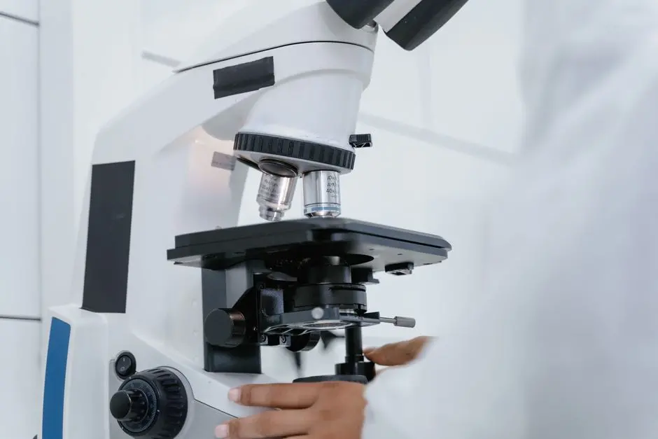
The quest for uncovering life's intricate mechanisms often begins under the lens of a microscope. Yet, selecting the precise instrument among the vast array of available technologies constitutes a pivotal decision, directly impacting the quality and relevance of your biological research. A misstep here can lead to wasted resources, ambiguous data, and missed discoveries. We confront this critical challenge head-on, empowering you with the strategic insights necessary to navigate the complex landscape of microscopy.
This authoritative resource meticulously dissects the core principles and practical considerations for choosing the optimal microscope tailored to your specific biological samples and experimental objectives. We dismantle the common pitfalls, illuminate the critical parameters of resolution, contrast, and sample compatibility, and provide a framework for informed decision-making. Prepare to unlock unprecedented clarity in your observations, enhancing everything from routine cell culture checks to groundbreaking subcellular dynamics. We delve into the distinctions that elevate standard visualization to powerful laboratory imaging methods for biological structures, ensuring your investment yields maximal scientific return.
Foundations of Microscope Selection: Defining Your Needs
Forging a successful microscopy strategy initiates with a ruthless self-assessment of your biological objectives. We must first define with surgical precision what we aim to observe. Consider the fundamental nature of your biological sample: Is it live or fixed? Are you examining whole organisms, tissue sections, individual cells, or subcellular organelles? Its inherent properties – transparency, thickness, size, and even its physiological state – dictate the initial filtering of potential microscope types. A thin bacterial smear demands a fundamentally different approach than a thick, opaque plant leaf or a beating heart in a live zebrafish embryo. This initial characterization is not merely a formality; it is the cornerstone upon which all subsequent decisions rest. We confront the reality that improper sample preparation can render even the most advanced microscope useless, emphasizing the need for robust protocols that preserve native morphology and function.
Next, delineate the specific biological information you seek to extract. Are you primarily interested in gross morphology, cellular processes in real-time, protein localization, or intricate ultrastructural details? This directly translates into requirements for resolution, magnification, and the need for dynamic imaging capabilities. Resolution, the ability to distinguish two closely spaced objects as separate, is often paramount. We recognize that magnification merely enlarges an image; without sufficient resolution, higher magnification only yields a larger blur. A typical light microscope might offer a maximum useful resolution of approximately 200 nanometers, governed by the Abbe diffraction limit, making structures below this threshold invisible. For visualizing intricate cellular architectures or individual protein complexes, alternative strategies are required. Conversely, for macroscopic dissections or observing large tissues, a stereo microscope offers ample working distance and a wide field of view, where high resolution is less critical. Neglecting these initial, critical questions is a common misstep, leading to suboptimal instrument choices and research bottlenecks. We empower researchers to articulate these needs clearly, ensuring alignment between biological question and technological capability and optimizing every step of the imaging workflow.
Demystifying Light Microscopy: Applications and Limitations
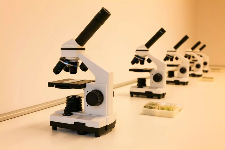
Light microscopy remains the workhorse of biological laboratories, offering unparalleled versatility and accessibility for a vast spectrum of applications. We navigate its diverse modalities, each engineered to address specific visualization challenges. Brightfield microscopy, the most fundamental form, illuminates the sample directly with white light. It excels with stained specimens, where differential absorption of light creates contrast, making cells and tissues readily visible. However, for unstained, living biological samples, which are largely transparent, brightfield offers limited contrast, rendering cellular structures indistinct. This is a crucial distinction: attempting to image live, unstained cells with brightfield often proves unproductive.
To overcome this, we deploy techniques that exploit differences in refractive index. Phase Contrast microscopy transforms subtle phase shifts in light passing through a transparent sample into amplitude differences, generating high-contrast images of unstained, living cells. It is indispensable for observing cell morphology, movement, and division without the need for toxic stains. Its primary drawback lies in the halo effect, which can obscure fine details. Differential Interference Contrast (DIC) microscopy, or Nomarski microscopy, represents a significant advancement. It utilizes polarized light and prisms to create an optical path difference across the sample, generating images with a pseudo-three-dimensional relief effect. DIC provides excellent resolution and contrast for live, unstained cells, often surpassing phase contrast in its ability to reveal intricate subcellular structures and reduce halo artifacts. We recognize that while these techniques are foundational for observing cellular processes in real-time, their resolution remains constrained by the diffraction limit of light, approximately 200 nm laterally and 500-700 nm axially. For visualizing structures below this threshold, or for molecular specificity, we must transition to more advanced imaging platforms.
The Power of Fluorescence: Unveiling Molecular Dynamics
To transcend the limitations of simple light absorption and refractive index, we harness the unparalleled power of fluorescence microscopy, a technique that has revolutionized modern biological research by enabling molecular-specific visualization. The principle is elegant: fluorophores absorb light at a specific excitation wavelength and emit light at a longer, lower-energy emission wavelength. By attaching these fluorophores to specific molecules within a cell – via antibodies (immunofluorescence), genetic fusions (e.g., GFP-tagged proteins), or specific dyes – we can precisely pinpoint their location, quantity, and dynamic behavior.
Epifluorescence microscopy provides a wide field of view and high sensitivity, making it ideal for visualizing labeled cells or tissue sections. However, it collects light from the entire thickness of the sample, leading to out-of-focus blur, particularly in thicker specimens. This limitation necessitates a move towards optical sectioning techniques. Confocal microscopy, a cornerstone of advanced cellular imaging, overcomes this by using pinholes to reject out-of-focus light, delivering crisp, high-resolution optical sections. We construct stunning 3D reconstructions of cells and tissues by stacking these optical sections, revealing intricate spatial relationships previously obscured. Its capacity for live cell imaging, coupled with the ability to simultaneously detect multiple fluorophores, makes it indispensable for studying protein trafficking, cell signaling, and organelle dynamics.
Even more advanced, multi-photon microscopy, typically two-photon, utilizes infrared light for excitation. This minimizes phototoxicity and photobleaching, allowing for deep tissue imaging in living organisms (in vivo) with exceptional penetration and reduced scattering. While requiring specialized and often costly equipment, its application in developmental biology, neurobiology, and clinical diagnostics is transformative. However, we acknowledge inherent challenges: phototoxicity and photobleaching remain considerations, particularly during prolonged live imaging, demanding careful experimental design and fluorophore selection. Nonetheless, fluorescence microscopy remains the gold standard for dissecting the molecular machinery of life with unprecedented precision.
Advanced Techniques and Specialized Microscopes
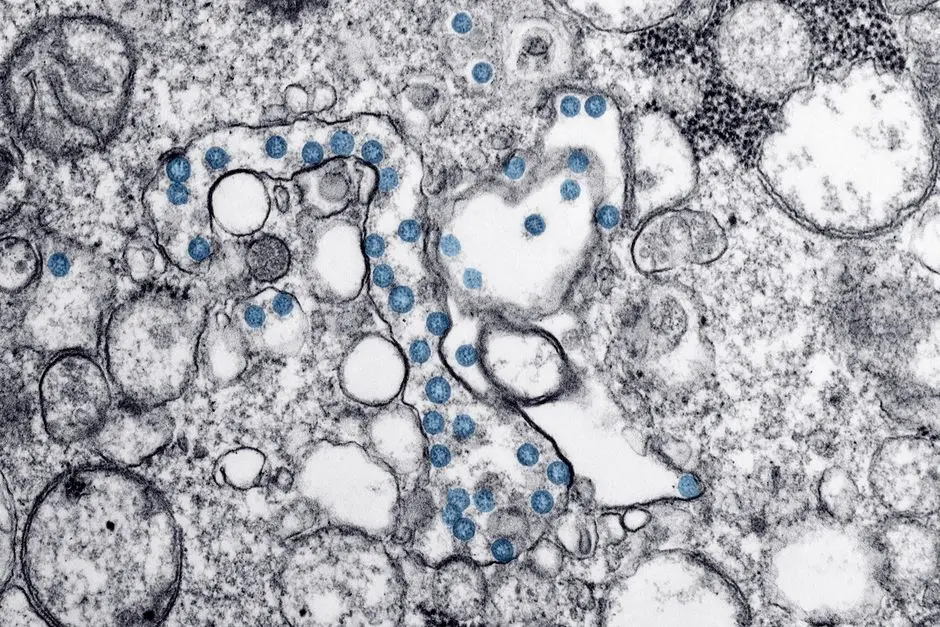
Beyond the realm of conventional fluorescence, we encounter a suite of highly specialized microscopy techniques engineered to tackle specific, challenging biological questions that push the boundaries of resolution, speed, and depth. When ultrastructural detail below the light diffraction limit becomes paramount – for visualizing viruses, protein complexes, or the intricate architecture of organelles – Electron Microscopy (EM) enters the arena. Transmission Electron Microscopy (TEM) delivers breathtaking 2D images of internal cellular structures, while Scanning Electron Microscopy (SEM) provides exquisite 3D surface topography. However, we must confront their significant limitations: EM requires extensive and often harsh sample preparation (fixation, dehydration, heavy metal staining, sectioning), rendering live imaging impossible. It is a powerful tool for fixed, non-dynamic snapshots of cellular architecture.
To break the diffraction barrier of light microscopy and achieve nanometer-scale resolution in live or fixed samples, we deploy Super-Resolution Microscopy techniques. Methods like STED (Stimulated Emission Depletion), PALM (Photoactivated Localization Microscopy), and STORM (Stochastic Optical Reconstruction Microscopy) optically overcome the diffraction limit, pushing resolution down to tens of nanometers. These techniques enable us to visualize individual proteins, molecular clusters, and intricate membrane structures with unprecedented clarity. While technically demanding and computationally intensive, they are indispensable for dissecting molecular organization within cells. For prolonged, gentle 3D imaging of large, delicate living samples like embryos or organoids, Light Sheet Fluorescence Microscopy (LSFM), or Selective Plane Illumination Microscopy (SPIM), stands as a revolutionary approach. It illuminates the sample with a thin sheet of light from the side, minimizing phototoxicity and photobleaching, allowing us to observe developmental processes over extended periods with minimal perturbation. These advanced microscopes represent significant investments in both capital and expertise, demanding a clear justification based on the biological question at hand. We leverage these tools to drive biological understanding where conventional methods fail.
Strategic Acquisition: Budget, Maintenance, and Future-Proofing
The selection of a microscope extends beyond its technical specifications; it encompasses a comprehensive strategic acquisition plan that factors in long-term viability and operational efficiency. We recognize that instrumentation represents a significant investment, often ranging from thousands for basic light microscopes to hundreds of thousands or even millions for advanced confocal or super-resolution systems. A critical error involves solely focusing on the initial purchase price. We meticulously calculate the Total Cost of Ownership (TCO), which includes not only the instrument itself but also essential accessories (objectives, cameras, software licenses), ongoing consumables (immersion oil, reagents, slides), and crucially, maintenance contracts. A robust service agreement safeguards your investment and minimizes downtime, a common oversight in budget planning.
Beyond direct costs, consider the logistical and human elements. What are the spatial and environmental requirements? Advanced microscopes often demand vibration isolation tables, dedicated darkrooms, stable temperature control, and specialized utility connections. Neglecting these environmental factors compromises imaging quality. Crucially, we evaluate the availability of vendor support, technical service, and comprehensive user training. A sophisticated microscope without skilled operators is an expensive ornament. We advocate for continuous training programs and the establishment of internal expertise to maximize instrument utility. Furthermore, consider the modularity and upgrade path of the system. Can you add new modalities (e.g., live cell incubation, TIRF, FRAP) as your research evolves? Future-proofing your investment ensures its relevance for years to come.
Finally, explore collaborative opportunities. Accessing high-end instruments through institutional core facilities often provides state-of-the-art technology, expert support, and cost-sharing benefits, democratizing access to cutting-edge microscopy. We actively champion leveraging these shared resources. Avoid the pitfall of purchasing features you may never fully utilize; instead, align capabilities directly with current and anticipated research needs. By adopting this strategic, holistic approach, we transform a significant capital expenditure into a powerful, sustainable engine for biological discovery, optimizing every facet of your imaging infrastructure.
Key Takeaways
Define Your Biological Question First
Before selecting any microscope, precisely articulate your experimental goals: sample type (live/fixed, transparency, size), desired information (morphology, dynamics, molecular localization), and essential resolution/magnification. This foundational step prevents costly misalignments.
Choose Contrast Techniques Wisely
For unstained, transparent live samples, deploy phase contrast or DIC microscopy to generate contrast from refractive index differences, enabling observation of morphology and movement without toxic stains. Brightfield is best for stained, fixed samples.
Leverage Fluorescence for Molecular Specificity
Fluorescence microscopy (epifluorescence, confocal, multi-photon) offers unparalleled specificity for visualizing target molecules via fluorophore labeling. Confocal excels in 3D imaging of thicker samples, while multi-photon allows deep tissue imaging with reduced phototoxicity.
Recognize Advanced Techniques for Extreme Resolution or Conditions
Electron microscopy (TEM/SEM) is indispensable for ultrastructural details beyond the light diffraction limit but requires fixed samples. Super-resolution methods (STED, PALM/STORM) optically break the diffraction barrier for nanometer precision. Light sheet microscopy enables gentle, long-term 3D live imaging.
Plan for Total Cost of Ownership and Long-Term Viability
Your acquisition strategy must include instrument cost, accessories, consumables, maintenance contracts, vendor support, user training, and environmental requirements. Consider modularity and core facility access to optimize investment and future-proof your research capabilities.
FAQ
-
What is the primary difference between resolution and magnification?
Resolution defines the ability to distinguish two separate entities as distinct, measured in nanometers, and is crucial for discerning fine details. Magnification, conversely, merely enlarges the image. High magnification without sufficient resolution only produces a larger, blurry image. We prioritize resolution as the fundamental metric for extracting meaningful biological information. -
When should I consider a confocal microscope over a standard epifluorescence microscope?
We advocate for confocal microscopy when imaging thicker samples (e.g., tissue sections, organoids, live embryos) or when precise 3D reconstruction is required. Confocal microscopes use pinholes to reject out-of-focus light, yielding significantly sharper optical sections and superior signal-to-noise ratios compared to epifluorescence, which collects light from the entire sample depth, leading to blur. -
Are there alternatives to fluorescent labeling for live cell imaging?
Absolutely. For live, unstained biological samples, we frequently deploy phase contrast and Differential Interference Contrast (DIC) microscopy. These techniques generate contrast by exploiting differences in refractive index within the sample, allowing visualization of cellular morphology and dynamics without the need for potentially phototoxic fluorescent probes. -
What crucial factors should I consider beyond the microscope's initial purchase price?
We emphasize calculating the Total Cost of Ownership (TCO), which extends beyond the instrument itself. Factor in the cost of high-quality objectives, specialized cameras, software licenses, ongoing consumables, and critically, a robust maintenance contract. Additionally, evaluate vendor support, training requirements, and the long-term upgradeability of the system to future-proof your investment.