
> Laboratory Techniques > Microscopy and Cellular Imaging > Unlock Cellular Secrets: Exploring Main Light Microscopy Types
Unlock Cellular Secrets: Exploring Main Light Microscopy Types
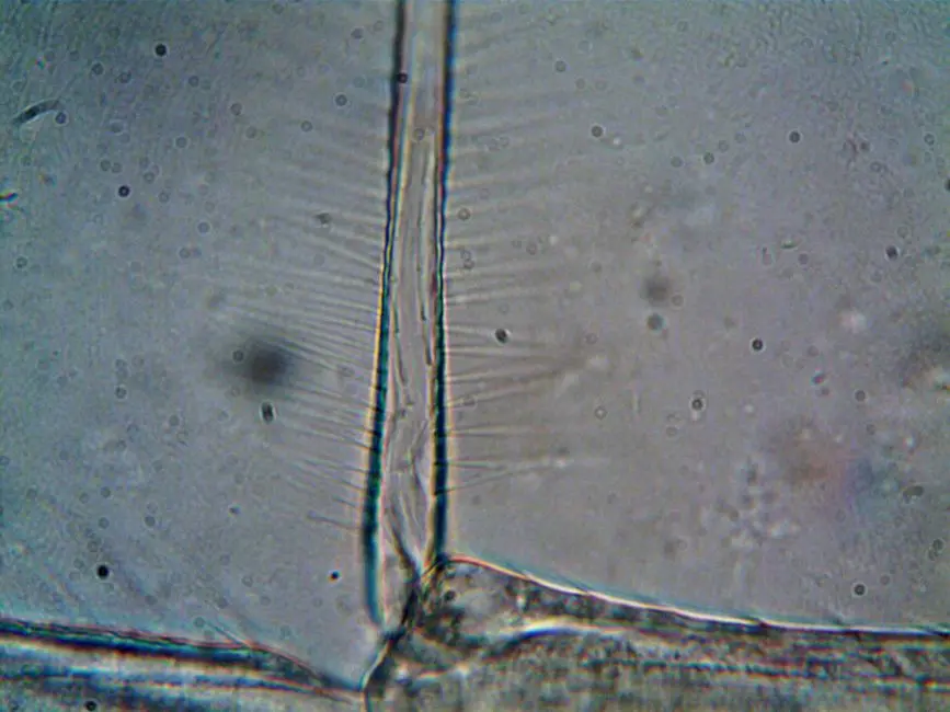
We forge knowledge by scrutinizing the invisible, and at the heart of this pursuit lies optical microscopy. This foundational technology remains the primary instrument for deciphering the architecture and dynamics of living cells. However, the plethora of available techniques presents a strategic challenge: how to choose the optimal optical tool to extract biological truth from our samples?
This article is not a mere directory; it is your surgical roadmap to navigating the complex landscape of major optical microscopy methods. We dissect the fundamental principles, targeted applications, and common pitfalls, transforming theory into tangible practice. Prepare to unlock unprecedented insights into microscopic life, exploring how each technique reveals unique facets. For a comprehensive understanding of biological imaging methods, feel free to consult our resource on laboratory imaging methods for biological structures. We equip you with the essential knowledge to propel your discoveries. Let's forge the next generation of cellular explorers together!
Illuminating the Invisible: Foundations of Light Microscopy
We begin our exploration by laying the groundwork for optical microscopy, an unwavering pillar of modern biology. This ingenious system utilizes visible light and a complex interplay of lenses to magnify the perception of structures too small for the naked eye. The fundamental principle relies on light's ability to interact with the sample, enabling the detection of specific contrasts or emissions. Two key concepts govern our ability to observe the microscopic world: resolution and magnification.
Magnification is the enlargement of the image, making it visible. Whereas resolution, the true cornerstone of our observational power, defines the ability to distinguish two distinct and closely spaced points. It is inherently limited by the wavelength of light used (the Abbe limit) and the numerical aperture (NA) of the objectives. A higher NA and a shorter wavelength allow for better resolution. We employ a precise optical path: the light source illuminates the sample through a condenser, the objective collects the transmitted or reflected light, and the eyepiece (or camera) projects the final image. Mastering these components is the first step to unlocking cellular secrets.
The strategic selection of objectives is crucial. Each objective possesses a specific magnification power and numerical aperture. A 100x objective with an NA of 1.4, for instance, offers superior resolution to a 100x with an NA of 1.25. We rely on plan-achromatic or plan-apochromatic objectives for flat images free from chromatic aberration, thereby ensuring the visual integrity of our discoveries. Understanding this synergy between light, lenses, and sample is not only essential but is the lever that allows us to propel our analyses towards unparalleled precision.

Bright-Field Microscopy: The Unveiling Baseline
Bright-field microscopy is the fundamental technique, the first gateway into the microscopic world that we have opened. Its principle is elegantly simple: light passes through the specimen, and variations in absorption or scattering by different structures create contrast. The denser or more colored the specimen, the more light it absorbs, appearing darker against a bright background.
We primarily use this method for examining fixed and stained specimens, such as histological sections, blood smears, or bacterial cultures. Staining is a key step, as it highlights specific structures or cell types that would otherwise be transparent. Stains like hematoxylin and eosin (H&E) in histology, or Gram stain in microbiology, are tools we use to reveal essential morphological details.
Despite its simplicity and relatively low cost, bright-field microscopy has significant limitations. Its main drawback is the low intrinsic contrast of unstained or living specimens. Living cells, largely composed of water, do not offer sufficient absorption variations to be clearly distinguished. This is where the major challenge lies: observing dynamic processes without altering cellular physiology. To ensure the quality of our observations, Köhler illumination is an indispensable practice. It optimizes illumination by ensuring uniform and maximal lighting of the field of view, while controlling the aperture diaphragm to balance contrast and resolution. Poorly adjusted Köhler illumination leads to low-quality images, directly affecting the validity of our interpretations. Let's avoid common errors: over or under-staining, air bubbles between slide and coverslip, or a dirty objective lens severely compromise image integrity. We demand meticulous preparation for every observation.
Sculpting Contrast: Phase Contrast and DIC Microscopy
To overcome the limitations of the brightfield and observe living, unstained specimens with high contrast, we employ more advanced optical techniques. Phase contrast microscopy and differential interference contrast (DIC) microscopy are our instruments for visualizing subtle differences in refractive index within cells, transforming them into variations in light intensity.
Phase contrast, a major innovation by Nobel laureate Frits Zernike, converts light phase shifts (caused by differences in sample thickness or refractive index) into amplitude variations, and thus brightness. We utilize an annular diaphragm in the condenser and a corresponding phase plate in the objective. Undiffracted light passes through a region of the phase plate that modifies its amplitude and shifts its phase by a quarter wavelength. Light diffracted by the specimen bypasses this region. Constructive or destructive interference between these two types of light generates contrast. This technique is invaluable for imaging live cells in culture, visualizing organelles without staining, and studying cell motility. Its advantage lies in its non-invasiveness and its ability to reveal subtle details. However, we must manage the "halo effect," a bright artifact around dense structures, which can obscure certain fine details.
DIC (Nomarski) microscopy takes this a step further. It uses polarized light and Wollaston prisms to split light into two orthogonally polarized rays, slightly offset from each other. These rays traverse the specimen at adjacent points, where they experience different phase delays based on the refractive index gradient. Upon recombination, these rays interfere to create an image with a distinctive pseudo-three-dimensional appearance, reminiscent of a bas-relief. DIC offers exceptional resolution and excellent optical sectioning, eliminating the halo effect of phase contrast. We apply it for high-resolution observations of live cells, flagella, cilia, and embryos. The higher cost and the need for specific objectives are factors to consider, but the clarity and richness of information that DIC provides often justify this strategic investment.
Illuminating Specificity: Fluorescence Microscopy
Fluorescence microscopy represents a revolution for biological observation, allowing us to visualize not generic shapes, but specific molecules within cells and tissues. Its principle relies on the ability of certain compounds, fluorophores, to absorb light at a specific wavelength (excitation) and then emit light at a longer wavelength (emission). This shift, known as Stokes shift, is the key to the specificity of this technique.
We use a specialized optical system: a powerful light source (arc lamp or laser) excites the fluorophores. Excitation filters select the appropriate wavelength, a dichroic mirror deflects the excitation light towards the sample while allowing the emission light to pass through, and emission filters transmit only the emitted fluorescent light. This setup almost entirely eliminates excitation light, leaving us with a remarkably clear signal solely from the labeled structures.
The applications of fluorescence microscopy are vast and powerful. We utilize it for the localization of specific proteins via immunofluorescence (using antibodies coupled to fluorophores) or the expression of fusion proteins with Green Fluorescent Protein (GFP) and its variants. It allows us to track dynamic processes in real-time in living cells, study protein-protein interactions, and quantify gene expression. The major advantage is the extreme sensitivity and molecular specificity, providing us with information inaccessible by other methods.
However, fluorescence is not without its challenges. Photobleaching (irreversible degradation of fluorophores under light excitation) and phototoxicity (cellular damage caused by light and generated free radicals) are obstacles we must overcome, by optimizing exposure parameters and choosing robust fluorophores. Autofluorescence from samples can also mask the signal of interest. To counter these limitations, we meticulously select fluorophores with appropriate excitation and emission spectra, optimize filter sets, and use anti-photobleaching mounting media. We control these parameters to ensure the robustness and reliability of our data.
Beyond 2D: Confocal and Super-Resolution Microscopy
To transcend the limitations of 2D imaging and the penetration depth of previous techniques, we turn to more sophisticated approaches. Confocal Laser Scanning Microscopy (CLSM) is a pivotal technique for 3D imaging and thick specimens, while super-resolution microscopy boldly pushes the diffraction limit.
Confocal microscopy operates on a principle of optical sectioning. Unlike wide-field microscopy where the entire specimen is illuminated simultaneously, confocal microscopy uses a laser to scan the specimen point by point. The light emitted from this point is collected and passes through a pinhole located in a plane conjugate to the sample's focal point. This pinhole is strategically placed to reject light originating from planes above and below the focal plane, significantly reducing blur and background noise. By digitally reconstructing these optical "slices," we forge a high-resolution 3D image. We apply this for imaging thick tissues, precise colocalization of molecules, and in-depth cellular dynamics studies. The advantages are clear: improved axial resolution, enhanced signal-to-noise ratio, and the ability to construct complex three-dimensional models. Limitations include slower acquisition times and potentially higher phototoxicity due to laser intensity, demanding rigorous optimization of acquisition parameters from us.
Going beyond Abbe's diffraction limit, super-resolution microscopy represents the pinnacle of optical imaging, allowing us to visualize structures with nanometer precision. Techniques such as STED (Stimulated Emission Depletion), PALM (Photoactivated Localization Microscopy), and STORM (Stochastic Optical Reconstruction Microscopy) circumvent this physical limit by selectively controlling the activation or deactivation of fluorophores. STED, for instance, uses a second laser to deactivate fluorescence around the focal spot, thereby effectively shrinking the emission area. PALM and STORM localize individual fluorophores by randomly activating and then deactivating them, accumulating precise positions to reconstruct a final ultra-high-resolution image. These technologies have ushered in a new era in cell biology, offering us unprecedented access to the nanoscale organization of protein complexes and subcellular structures. Although complex and costly, their power of revelation is unmatched, transforming our understanding of life's fundamental processes.
Strategic Mastery: Selecting Your Optical Weaponry
Mastering the types of optical microscopy lies not only in understanding their principles but, crucially, in the strategic selection of the right tool for our specific biological question. Each technique is a different blade in our arsenal, and an expert knows when to sharpen which. We must evaluate several crucial factors when making this choice:
- Nature of the sample: Is it live or fixed? Transparent or opaque? Flat or thick? For live, transparent cells, phase contrast or DIC are favored. For fixed and stained tissues, brightfield is often sufficient. For dynamic processes or molecular localization, fluorescence is essential.
- Information sought: Do we want to observe general morphology, the localization of a specific molecule, the dynamics of a process, or a detailed 3D reconstruction?
- Required resolution: A broad overview (brightfield) or nanometric details (super-resolution)?
- Budget and accessibility: Advanced systems like confocal or super-resolution represent a significant investment.
Key Takeaways
Understanding Resolution vs. Magnification
Magnification enlarges the image, while Resolution dictates the ability to distinguish fine details. Resolution, limited by light's wavelength and numerical aperture (NA), is paramount for scientific discovery.
Bright-Field Microscopy: The Baseline
Ideal for fixed, stained samples. Relies on light absorption/scattering for contrast. Key limitation: poor contrast for live, unstained cells. Köhler illumination is vital for uniform, high-quality lighting and image clarity.
Enhanced Contrast for Live Imaging: Phase Contrast & DIC
- Phase Contrast: Converts refractive index variations into brightness changes, ideal for unstained live cells, but can produce halo artifacts.
- DIC (Nomarski): Uses polarized light for pseudo-3D relief based on refractive index gradients, offering high resolution and optical sectioning without halos, superior for detailed live-cell morphology.
Molecular Specificity: Fluorescence Microscopy
Uses fluorophores (excitation/emission) to visualize specific molecules. Powerful for immunofluorescence, GFP-tagging, and live-cell tracking. Challenges include photobleaching, phototoxicity, and autofluorescence, requiring careful experimental setup.
Advanced 3D & Nanoscale Imaging: Confocal & Super-Resolution
- Confocal Microscopy: Utilizes a pinhole to achieve optical sectioning and 3D reconstruction, reducing out-of-focus light for sharper images in thick samples.
- Super-Resolution Microscopy (STED, PALM, STORM): Overcomes the diffraction limit to achieve nanoscale resolution, revealing ultrastructural details previously inaccessible with light.
Strategic Selection & Future Outlook
Choosing the correct microscopy technique hinges on sample type (live/fixed), research question, desired resolution, and budget. Future trends include AI integration, correlative light and electron microscopy (CLEM), and novel fluorophores, continually expanding our visualization capabilities.
FAQ
-
What is the primary difference between resolution and magnification?
Magnification is the process of enlarging an object's apparent size, making it bigger. Resolution, however, defines the ability to distinguish two closely spaced objects as separate entities. A microscope can magnify an image many times, but if the resolution is poor, the magnified image will simply appear as a larger blur, without revealing finer details. Resolution is the true limiting factor in observing microscopic structures.
-
When should I choose phase-contrast over bright-field microscopy?
We decisively choose phase-contrast microscopy when observing unstained, live biological samples, such as cultured cells, bacteria, or tissue sections, without introducing toxic stains. Bright-field microscopy, while simpler, provides very poor contrast for transparent samples unless they are fixed and stained, a process that kills cells and can introduce artifacts. Phase contrast converts subtle differences in refractive index within the live sample into visible brightness variations, allowing us to study cell morphology, motility, and division in their natural, dynamic state.
-
What are the main limitations of fluorescence microscopy?
Fluorescence microscopy, while powerful for its specificity, faces several key limitations we must navigate. Foremost are photobleaching (the irreversible destruction of fluorophores upon prolonged light exposure) and phototoxicity (damage to live cells caused by the exciting light or reactive oxygen species generated). Additionally, autofluorescence from endogenous cellular components can obscure specific signals, leading to increased background noise. The need for specific fluorophores and filter sets also adds to experimental complexity and cost. We must proactively optimize our experimental conditions, selecting robust fluorophores and minimizing light exposure to mitigate these challenges.
-
Can light microscopy achieve nanoscale resolution?
Historically, conventional light microscopy was limited by the diffraction barrier to a resolution of approximately 200-250 nanometers. However, super-resolution microscopy techniques, such as STED, PALM, and STORM, have fundamentally overcome this barrier. These advanced methods allow us to achieve resolutions in the 20-50 nanometer range, effectively pushing light microscopy into the nanoscale realm. This breakthrough empowers us to visualize individual molecules and nanoscale cellular structures with unprecedented detail, revolutionizing our understanding of cellular architecture and function.
-
What is Köhler illumination and why is it important?
Köhler illumination is a critical technique for optimizing the illumination path in bright-field microscopy, ensuring we achieve the highest possible image quality. Its importance lies in producing uniform and bright illumination across the entire field of view, free from glare and artifacts. By properly adjusting the field diaphragm and the aperture diaphragm, we precisely control the light's path to the specimen. This process maximizes contrast and resolution simultaneously, while minimizing specimen heating and photobleaching, allowing us to capture images with optimal clarity and accuracy. It is a foundational practice for serious microscopy.