
> Laboratory Techniques > Microscopy and Cellular Imaging > Harness Optical Microscopy: Essential Lab Applications Explored
Harness Optical Microscopy: Essential Lab Applications Explored
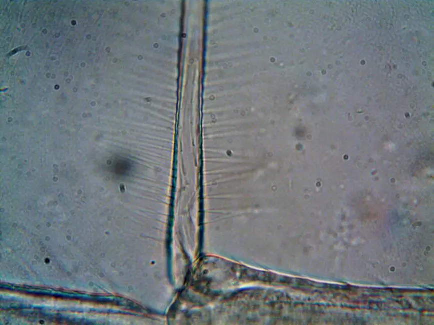
Let's unlock the incredible potential of the microscopic world. Microscopic optics is not just a technique; it is the cornerstone of modern biology, allowing us to visualize the invisible and decipher the fundamental mechanisms of life. From early cellular observations to dynamic subcellular analyses, its evolution is transforming our understanding. We harness its power to explore tissue structure, cellular dynamics, molecular interactions, and much more.
This article synthesizes the critical applications of optical microscopy in contemporary biology laboratories. We will dissect essential techniques, common pitfalls, and best practices that catalyze discovery. Prepare to forge technical mastery that will propel your research, deepening our exploration of advanced laboratory imaging methods for biological structures. We will equip you with strategies to optimize every experiment, ensuring reliable data and profound understanding.
Mastering the Fundamental Techniques of Optical Microscopy
We base our exploration on the pillars of optical microscopy, essential tools that remain indispensable in every biology laboratory. Bright-field microscopy, the most basic, reveals the morphology of cells and tissues through simple light absorption. It is ideal for stained specimens but has limited resolution, and unstained specimens appear almost transparent. We overcome this limitation with phase contrast microscopy and differential interference contrast (DIC) microscopy.
Phase contrast converts light phase shifts, induced by refractive index variations within the specimen, into variations in light intensity. This technique is perfect for observing unstained live cells, allowing us to discern organelles without altering their integrity. Its main drawback is halo artifact. DIC, on the other hand, uses Wollaston prisms to create contrast based on the refractive index gradient. It produces a very sharp, three-dimensional image, free from the halo of phase contrast, and is particularly useful for detailed morphological analysis and small intracellular movements.
Insider Tips for Optimization:
Green Light Filter: For phase contrast, a green light filter often enhances contrast.
Specimen Thickness: Keep specimens thin to maximize resolution and minimize optical aberrations, especially in DIC.
Numerical Aperture Setting: Always optimize the numerical aperture (NA) of both the objective and condenser to balance contrast and resolution. A higher NA improves resolution but can reduce depth of field.
We trigger precise observations by understanding these fundamental techniques and applying them judiciously, with each instrumentation choice being a strategic decision for the clarity of our data.
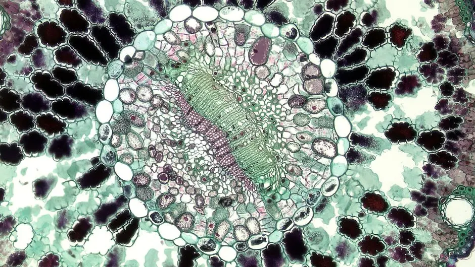
Illuminating Cellular Dynamics: Fluorescence and Confocal Imaging
To probe cellular mechanisms with molecular precision, we turn to fluorescence microscopy. This technique leverages the ability of certain molecules (fluorophores) to absorb light at a specific wavelength and emit it at another, longer wavelength. We utilize this property to selectively label specific structures or proteins within cells, often via fluorophore-conjugated antibodies (immunofluorescence) or intrinsic fluorescent proteins like GFP.
However, conventional fluorescence microscopes suffer from out-of-focus blur originating from planes above and below the focal plane. This is where confocal microscopy steps in, elegantly addressing this challenge. By employing a pinhole to reject non-focal light, the confocal microscope allows us to acquire thin optical sections of thick samples, thereby creating 3D reconstructions of unparalleled clarity. We optimize its use for colocalization studies, protein dynamics via FRAP (Fluorescence Recovery After Photobleaching), and FRET (Förster Resonance Energy Transfer), essential methods for analyzing molecular interactions.
Common Pitfalls to Avoid:
Photobleaching: Prolonged exposure to excitation light can degrade fluorophores. Let's minimize exposure time and laser power.
Autofluorescence: Biological tissues exhibit natural fluorescence. Let's use fluorophores with emission spectra distant from the sample's autofluorescence.
Image Saturation: Overexposure can saturate pixels, masking precious details. Let's meticulously adjust light intensity and detector gain.
We forge robust data by navigating the power of fluorescence and the precision of confocal imaging, transforming cellular imaging into a source of quantitative and dynamic information.
Quantifying Biological Processes: Image Analysis and Data Interpretation
Beyond simple visualization, our ultimate goal is quantification. Optical microscopy generates raw data that we must transform into actionable information. Image analysis is a critical step where we measure, segment, and interpret captured structures and events. Software such as ImageJ/FIJI, CellProfiler, and Imaris are our allies in extracting quantitative parameters like cell size, fluorescence intensity, organelle count, or the distance between two molecules.
Segmentation is often the first and most delicate step: we identify the boundaries of objects of interest. Accurate segmentation is the foundation of any reliable quantitative analysis. Subsequently, we perform intensity measurements to quantify protein expression or colocalization measurements to assess protein-protein interactions. For dynamic studies, we track objects over time to analyze their movement or morphological changes.
Best practices for analysis:
Rigorous controls: Always include positive and negative controls to validate our labeling and analysis methods.
Standardization: Apply standardized analysis protocols to all samples to minimize bias.
Objective thresholding: Use automatic or semi-automatic thresholding algorithms rather than subjective manual thresholding for maximum reproducibility.
Statistical validation: Do not settle for qualitative visualization; confirm our observations with robust statistical analyses.
We thus forge solid conclusions by transforming our images into numerical data, enabling a quantitative and reproducible understanding of biological phenomena.
Pioneers of Biology: Advanced Microscopy and Emerging Trends
As we push the boundaries of resolution and visualization, new optical microscopy techniques are emerging, revolutionizing our ability to explore the biological world. Super-resolution microscopy, including methods like STED (Stimulated Emission Depletion) and PALM/STORM (Photoactivated Localization Microscopy/Stochastic Optical Reconstruction Microscopy), has broken the optical diffraction limit, enabling us to visualize subcellular structures with nanometer resolution. We can now resolve protein aggregates, nuclear pores, and individual synapses, details that were previously inaccessible.
Another major advancement is Light-Sheet Microscopy (LSM). By illuminating a thin focal plane with a sheet of light and detecting fluorescence perpendicularly, we minimize phototoxicity and photobleaching, which is crucial for long-term imaging of live samples, such as developing embryos. LSM allows for rapid acquisition of volumetric 3D images with excellent sample integrity.
Future Trends and Opportunities:
Artificial Intelligence (AI): We are integrating AI for image enhancement (denoising, virtual super-resolution), automated segmentation, and phenotypic classification, accelerating and optimizing the analysis of massive datasets.
Correlative Microscopy: We combine optical and electron microscopy (CLEM) to locate rare events or structures with high ultrastructural resolution.
Miniaturization and Portability: The development of more compact and affordable microscopes is opening new avenues for imaging in the field or in clinical settings.
We are forging the future of biology by embracing these pioneering technologies, transforming once intractable questions into opportunities for unprecedented discovery.
Key Takeaways
Optical Foundations for Cell Biology
We master fundamental techniques such as brightfield, phase contrast, and DIC for visualizing live cells and tissues. These methods are crucial for morphological observation, with phase contrast and DIC offering vital information on unstained structures and cell dynamics without harming the sample. Careful optimization of sample thickness and numerical aperture is essential for accurate results.
Unlocking Molecular Interactions with Fluorescence
We leverage the power of fluorescence and confocal microscopy to illuminate subcellular structures and molecular dynamics. Fluorescence allows us to label specific molecules, while confocal microscopy provides us with 3D optical resolution and reduced out-of-focus blur. We avoid photobleaching and autofluorescence to ensure data integrity, and employ techniques such as FRAP and FRET for in-depth protein interaction analyses.
Quantify Our Biological Discoveries
Quantification of microscopic images is a necessity. We utilize analysis software for segmentation, intensity measurements, and colocalization, transforming raw images into actionable digital data. The adoption of rigorous controls, standardized protocols, and objective thresholding ensures the reliability and reproducibility of our analyses, enabling us to forge statistically robust conclusions.
Navigating the Future of Biological Imaging
We embrace innovations such as super-resolution microscopy (STED, PALM/STORM) and light-sheet microscopy (LSM) to overcome the limitations of optical resolution and minimize phototoxicity. These techniques open windows for us into nanometer-scale details and long-term volumetric imaging. The integration of artificial intelligence for image enhancement and analysis represents an exciting frontier, optimizing our ability to extract complex information from our data.
FAQ
-
What optical microscopy technique is best for observing live cells with high contrast?
To observe unstained living cells with high contrast, we favor phase contrast microscopy or differential interference contrast (DIC) microscopy. Phase contrast is simple to use and very effective for intracellular details, while DIC offers superior optical resolution and a halo-free, quasi-3D appearance, ideal for finer morphological analyses. -
How to minimize photobleaching and phototoxicity in fluorescence microscopy?
We minimize photobleaching and phototoxicity through several key strategies:
Reduce excitation laser/light power: Use the lowest setting that provides sufficient signal.
Limit exposure time: Acquire images as quickly as possible.
Use photostable fluorophores: Choose fluorophores that are inherently more resistant to degradation.
Add anti-photobleaching agents: Incorporate reagents into our mounting buffers.
Light-sheet microscopy (LSM): For long-term live-cell imaging, LSM is superior because it only illuminates a thin focal plane.
-
What are the main challenges of quantitative image analysis in microscopy?
Quantitative image analysis presents several challenges that we must overcome:
Precise segmentation: Separating objects of interest from the background is often complex, especially for irregular or low-contrast structures.
Noise and artifacts: Images may contain noise or artifacts that distort measurements.
Inter-sample variability: Variations in preparation or acquisition can introduce biases.
Protocol standardization: Ensuring the reproducibility and objectivity of the analysis is essential and requires rigorous protocols.
Software complexity: Mastering advanced image analysis software requires expertise and training.
-
How does super-resolution microscopy differ from conventional optical microscopy?
Super-resolution microscopy (such as STED, PALM/STORM) overcomes the optical diffraction limit, a physical barrier that restricts the resolution of conventional optical microscopes to approximately 200-250 nm. While conventional microscopy (fluorescence, confocal) allows us to visualize cells and organelles, super-resolution propels us into the nanometric realm, revealing subcellular and molecular details with a precision of up to 20-30 nm, opening new windows into the fine organization of proteins and structures within cells.