
> Laboratory Techniques > Microscopy and Cellular Imaging > Unlocking Cellular Secrets: Brightfield vs. Phase Contrast Microscopy
Unlocking Cellular Secrets: Brightfield vs. Phase Contrast Microscopy
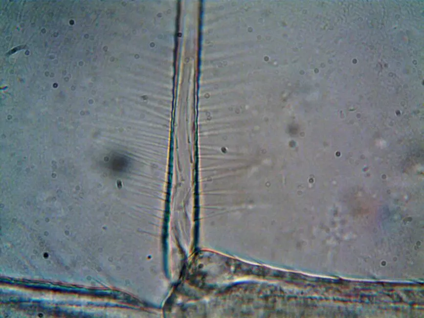
In the vibrant tapestry of biological exploration, the microscope stands as our indispensable eye, peering into realms invisible to the naked eye. Yet, not all microscopic visions are created equal. The fundamental challenge for any biologist is rendering the transparent, often colorless, architecture of living cells visible without compromising their integrity.
We embark on a strategic journey to dissect and master two foundational pillars of light microscopy: brightfield and phase contrast. These techniques, while both leveraging visible light, employ distinct mechanisms to generate crucial contrast, each with its unique arsenal of strengths and limitations. This article will equip you with the expert knowledge to decisively choose the optimal tool for your specific biological query, transforming ambiguity into clarity.
Forge an unshakeable understanding of their underlying principles, identify common pitfalls, and embrace advanced strategies to maximize your observational power. From stained tissue sections to live, unstained cellular dynamics, we demystify the science, ensuring you extract maximum value from every microscopic observation. Prepare to elevate your understanding of laboratory imaging methods for biological structures and revolutionize your approach to cellular visualization. Our objective: to arm you with the precision required to unveil the intricate ballet of life at its most fundamental level.
The Foundational Gaze: Understanding Brightfield Microscopy
Brightfield microscopy serves as the bedrock of biological imaging, renowned for its straightforward approach and universal applicability. At its core, the technique illuminates a sample with visible light, collecting the light that passes directly through it or is scattered. The resulting image exhibits contrast based primarily on the absorption of light by the specimen's components. Areas that absorb or scatter more light appear darker against a bright background, hence the 'brightfield' designation.
This method shines brightest when dealing with specimens that naturally possess pigments, like photosynthetic organisms, or more commonly, those that have been artificially stained. Staining protocols, such as Hematoxylin and Eosin (H&E) for tissue sections or Gram staining for bacteria, are critical for brightfield's efficacy. These dyes selectively bind to specific cellular structures, enhancing their light absorption properties and rendering them visible. Without staining, many biological samples, particularly living cells, appear almost entirely transparent, offering minimal information due to their similar refractive indices to the surrounding medium.
The ease of use and cost-effectiveness of brightfield microscopes make them ubiquitous in educational settings and clinical laboratories for routine analysis. However, their reliance on staining introduces a significant limitation: the staining process often kills or alters the specimen, precluding the observation of live cellular dynamics. We must recognize brightfield's power in revealing fixed, structural details, while acknowledging its inherent blind spots when faced with the delicate dance of living, unstained biology.
Mastering the Brightfield Technique: Principles, Applications, and Limitations
To truly master brightfield microscopy, we must delve into its operational nuances. The critical components include a light source, a condenser to focus light onto the specimen, the objective lens for magnification, and the eyepiece for viewing. Optimal image quality hinges on meticulous illumination setup, primarily Köhler illumination, which ensures even, bright, and high-contrast illumination across the field of view. Proper adjustment of the condenser aperture is paramount; balancing resolution and contrast prevents excessive glare that can obscure fine details.
Applications:
- Histology and Pathology: Examining stained tissue sections for diagnostic purposes (e.g., cancer detection, tissue abnormalities).
- Microbiology: Observing stained bacterial smears to identify morphology and Gram stain reactions.
- Parasitology: Identifying stained parasites in blood or fecal samples.
- Basic Cell Biology: Analyzing fixed and stained cell cultures to study general morphology and subcellular structures.
Limitations:
- Lack of Contrast for Unstained Samples: This is the paramount drawback. Living cells and many thin biological materials are transparent, making them invisible without significant chemical intervention.
- Artifacts from Staining: Chemical stains can introduce artifacts, alter cellular structures, or kill living specimens, making it impossible to observe natural, dynamic processes.
- Limited Depth of Field: For thicker specimens, only a thin plane is in focus at any given time, requiring constant refocusing.
- Phototoxicity: Although less pronounced than with fluorescent techniques, prolonged exposure to high-intensity light can still damage sensitive specimens, especially if they are stained.
We must strategically deploy brightfield when fixed, stained samples are our focus, leveraging its simplicity and proven utility for structural analysis.
Phase Contrast Microscopy: Illuminating the Invisible Dynamics of Life
When the challenge shifts from fixed structures to the dynamic processes within living, unstained cells, phase contrast microscopy emerges as the indispensable solution. Pioneered by Frits Zernike, this technique ingeniously converts subtle phase shifts in light passing through a specimen—caused by variations in refractive index and thickness—into detectable differences in brightness (amplitude). Most biological specimens are largely transparent because their components have similar refractive indices to water, making them indistinguishable under brightfield.
The core principle lies in manipulating the phase of light waves. A phase contrast microscope utilizes two key components: an annular diaphragm in the condenser and a phase plate positioned within the objective lens. The annular diaphragm creates a hollow cone of light, directing it through the specimen. Undiffracted light (the 'surround' light that passes through the specimen without being scattered) passes directly through a specific ring on the phase plate. Diffracted light (light scattered by the specimen's internal structures due to refractive index differences) passes through other parts of the phase plate.
Critically, the phase plate introduces a 1/4 wavelength phase shift to the undiffracted light and often attenuates its amplitude. When the phase-shifted undiffracted light recombines with the diffracted light, constructive and destructive interference occurs. Areas where light waves reinforce appear brighter, and areas where they cancel out appear darker, revealing the internal architecture of otherwise transparent cells. This conversion of invisible phase differences into visible amplitude differences allows us to visualize organelles, membranes, and cellular movements in real-time, without invasive staining.
Optimizing Phase Contrast: Principles, Components, and the Halo Effect
Successful phase contrast imaging demands a precise interplay of its specialized optical components. The annular diaphragm, inserted into the condenser, restricts the illuminating light to an annulus, ensuring that only a specific portion of the light interacts with the phase plate. The phase plate, located at the back focal plane of the objective lens, is a crucial component with a precisely machined ring that matches the annulus of the condenser diaphragm. This ring on the phase plate is designed to retard the phase of the undiffracted light by approximately 1/4 wavelength relative to the diffracted light, and often also to absorb a portion of the undiffracted light to balance intensity.
The alignment between the annular diaphragm and the phase plate is critical. Misalignment results in suboptimal contrast and image quality. Most phase contrast systems include a centering telescope or Bertrand lens to facilitate this alignment, ensuring that the condenser annulus is perfectly superimposed with the phase ring in the objective's back focal plane.
A characteristic feature of phase contrast images is the 'halo effect' – a bright ring often seen around the perimeter of dense structures. This phenomenon arises because some diffracted light inevitably passes through the phase ring, and some undiffracted light passes outside it, leading to slight phase mismatches at interfaces. While a minor drawback, the halo effect rarely impedes the interpretation of critical cellular details. Proper objective numerical aperture and condenser adjustments can minimize its prominence. We conquer the challenge of observing transparent specimens by meticulously orchestrating these optical interactions, unlocking a world of dynamic biological processes previously veiled in invisibility.
Strategic Selection: Brightfield vs. Phase Contrast – A Decision Framework
Our ultimate goal is not merely understanding, but strategic deployment. Choosing between brightfield and phase contrast microscopy hinges directly on the nature of your specimen and your research objectives. We forge a clear decision framework:
Brightfield Microscopy: The Structural Cartographer
- Best for: Fixed, stained specimens. Histological sections, bacterial smears, fixed cell cultures, samples with natural pigmentation.
- Advantages: Simple, cost-effective, excellent for visualizing morphology and spatial relationships after staining, provides color information from dyes.
- Limitations: Destructive staining, no observation of live dynamics, poor contrast for unstained transparent samples.
- Key Use Case: Diagnostic pathology, identifying specific cell types or pathogens in prepared slides.
Phase Contrast Microscopy: The Dynamic Narrator
- Best for: Living, unstained cells and thin, transparent biological materials (e.g., protozoa, cell cultures, fresh tissue sections).
- Advantages: Visualizes internal structures without staining, allows real-time observation of cellular processes (motility, division, organelle movement), non-invasive.
- Limitations: Grayscale images, 'halo effect,' requires specialized optics (annular diaphragm, phase plate), cannot typically be used with thick specimens, specific objective-condenser matching.
- Key Use Case: Studying cell division, phagocytosis, cell migration, drug effects on live cells.
Insider Tip: For initial screening of a live cell culture, phase contrast is often superior. If specific subcellular details require higher resolution or fluorescent markers, other advanced techniques might be necessary, but phase contrast provides the foundational live view. We must consider the entire experimental workflow—from sample preparation to data analysis—to make the most impactful choice for our biological inquiry.
Key Takeaways
Brightfield Microscopy: The Foundation for Fixed Samples
Principle: Relies on light absorption/scattering; creates contrast for naturally pigmented or stained specimens. Most living, unstained cells are transparent.
Applications: Histology, pathology (stained tissues), microbiology (stained bacteria), fixed cell cultures.
Limitations: Staining kills/alters cells, no live observation, poor contrast for unstained samples.
Key Feature: Simple, cost-effective, provides structural details with color.
Phase Contrast Microscopy: Unveiling Live Cellular Dynamics
Principle: Converts subtle phase shifts (due to refractive index differences) into visible brightness variations. Uses an annular diaphragm and a phase plate.
Applications: Live cell imaging, observing cell division, motility, organelle movement in unstained specimens.
Advantages: Non-invasive, real-time observation of dynamic processes, no need for staining.
Limitations: Grayscale images, 'halo effect,' requires specialized optics and precise alignment.
Key Feature: Enables visualization of transparent, living cells and their internal structures.
Strategic Choice: Aligning Technique with Research Goals
Brightfield: Opt for fixed, stained samples when structural and morphological details are paramount and live observation is not required.
Phase Contrast: Choose for living, unstained specimens to study dynamic cellular behaviors and processes without introducing artifacts from dyes.
Expert Tip: Always consider your sample's state (live/fixed) and your observational objective (structure/dynamics) to make the most informed decision.
FAQ
-
Why do unstained living cells appear transparent under brightfield microscopy?
Unstained living cells are largely transparent because their internal components (cytoplasm, organelles) have refractive indices very similar to their aqueous environment. Brightfield microscopy relies on differences in light absorption or scattering to create contrast. Since transparent cells absorb and scatter very little light, there's insufficient contrast to distinguish structures, rendering them virtually invisible against the bright background.
-
What is the 'halo effect' in phase contrast microscopy and how does it impact observations?
The 'halo effect' is a characteristic optical artifact in phase contrast images, appearing as a bright ring around dense structures (e.g., cell boundaries, organelles). It arises from some diffracted light passing through the undiffracted light's pathway on the phase plate, and vice versa. While it can slightly obscure very fine details at interfaces, it generally does not impede the visualization of overall cellular morphology or dynamic processes. It's a trade-off for the ability to see unstained living cells.
-
Can I use phase contrast objectives with a standard brightfield condenser?
No, phase contrast objectives require a specific phase contrast condenser equipped with an annular diaphragm that matches the phase plate in the objective. A standard brightfield condenser produces a solid cone of light, which would not interact correctly with the phase plate, resulting in poor or no phase contrast effect. Proper alignment between the condenser's annulus and the objective's phase plate is essential for generating optimal contrast.
-
When would brightfield microscopy be preferred over phase contrast, despite its limitations?
Brightfield microscopy is preferred when examining fixed and stained specimens, especially for diagnostic purposes where specific cellular structures or pathogens are highlighted by dyes. It's also chosen for samples with natural pigmentation. While it cannot observe live dynamics, its simplicity, cost-effectiveness, and the ability to add color information through stains make it invaluable for structural analysis in histology, pathology, and certain microbiological applications where cell viability is not a concern.