
> Laboratory Techniques > Microscopy and Cellular Imaging > Forge Your Live-Cell Microscopy Setup: The Essential Arsenal
Forge Your Live-Cell Microscopy Setup: The Essential Arsenal
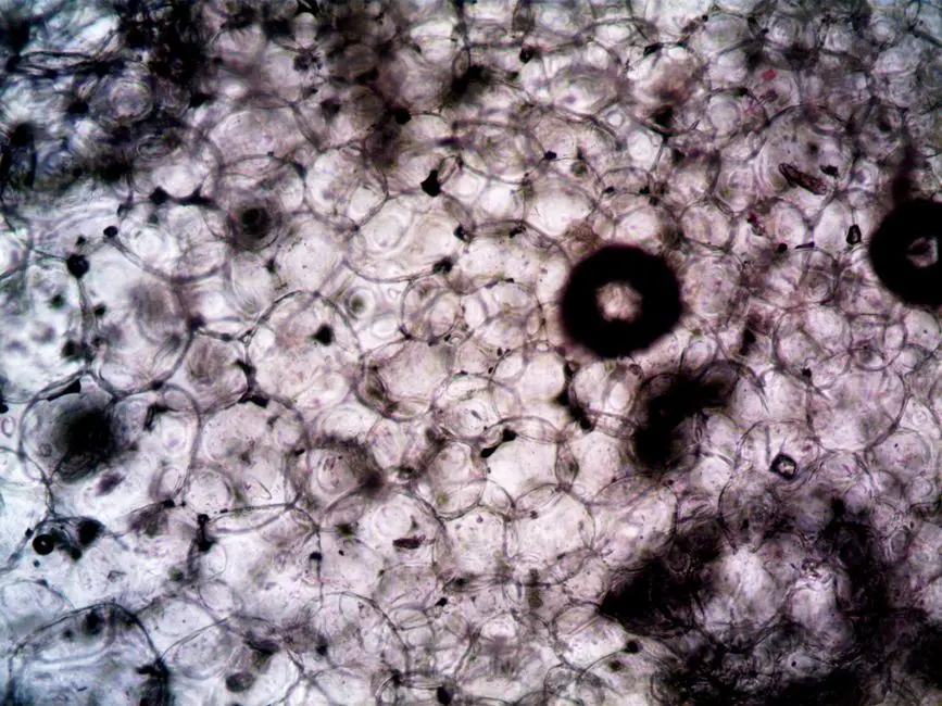
Unveiling the dynamic processes within living cells represents a frontier in biological research, driving breakthroughs from neurobiology to oncology. To truly comprehend cellular mechanisms, static images simply do not suffice; we demand real-time observation of cells interacting, moving, and responding. This pursuit necessitates a specialized toolkit, far beyond the scope of traditional fixed-cell imaging.
We are about to embark on a surgical exploration of the indispensable equipment that forms the backbone of any successful live-cell microscopy workflow. Mastering advanced laboratory imaging methods for biological structures is paramount, and live-cell microscopy stands as a cornerstone in this quest, empowering scientists to witness life unfold. We will dissect each critical component, from high-precision optical systems to sophisticated environmental controls and cutting-edge detectors, ensuring you forge a setup capable of capturing the most subtle biological events with unparalleled clarity and temporal resolution. Prepare to optimize your research potential by understanding the strategic choices that define state-of-the-art live-cell imaging.
The Core Foundation: Microscope Platforms and Advanced Optics
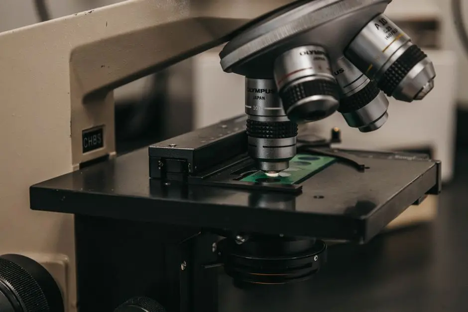
The bedrock of any live-cell microscopy setup is the microscope platform itself, predominantly an inverted microscope. This configuration allows objective lenses to be positioned beneath the specimen, leaving ample space above the stage for environmental control units, microfluidics, or micromanipulators. We prioritize a motorized stage for precise positioning, multi-point imaging, and the ability to revisit specific regions of interest over extended periods. Precision in movement is non-negotiable for reliable long-term observation.
Optics represent the eyes of our system. High-quality objective lenses are paramount. We must select objectives with a high Numerical Aperture (NA) for superior light collection and resolution, while also considering long working distances (WD) to accommodate dishes, chambers, and environmental control components. Air, water, or oil immersion options each present unique advantages. For live-cell work, water immersion objectives often strike an excellent balance, minimizing aberrations while remaining compatible with aqueous samples. Crucially, objectives must be corrected for temperature and media variations, often featuring correction collars to fine-tune optical performance and minimize spherical aberration caused by differing refractive indices of the sample medium and vessel. Neglecting these details degrades image quality, rendering dynamic events ambiguous. Investing in a robust, vibration-isolated optical table also significantly enhances stability, preventing focus drift and movement artifacts that compromise data integrity during prolonged imaging sessions.
Sustaining Life: Environmental Control and Specimen Management
Maintaining physiological conditions for living cells throughout an experiment is as critical as the optics themselves. Even slight deviations in temperature, pH, or humidity can induce stress, alter cellular behavior, and ultimately invalidate results. We must integrate a comprehensive environmental control system. This typically includes a temperature control unit that precisely regulates the specimen chamber, maintaining a constant 37°C for mammalian cells. Beyond temperature, active CO2 gas controllers are essential to maintain the correct pH of the cell culture media, typically around pH 7.4. These systems often work in conjunction with humidity control modules to prevent evaporation from culture dishes, which can lead to changes in osmolarity and concentration of nutrients or drugs, rapidly compromising cell viability.
Various specimen chambers and incubators are available, ranging from simple heating inserts for multi-well plates to full-enclosure incubators that surround the entire microscope stage. The choice depends on the experiment's duration and complexity. For long-term time-lapse experiments, minimizing evaporation and maintaining sterility are paramount. We must consider specialized perfusion systems for delivering fresh media or reagents to cells, or microfluidic devices for highly controlled microenvironments. A common pitfall is focus drift due to thermal expansion and contraction; advanced systems integrate autofocus mechanisms (hardware-based or software-driven) to maintain consistent focal planes over hours or days, ensuring our dynamic data remains sharp and interpretable. Prioritizing robust environmental control elevates data quality and the biological relevance of our observations.
Capturing Light: Advanced Illumination and Detection Systems
The illumination source dictates how we visualize our cells, while the detector converts light into digital images. For live-cell microscopy, minimizing light exposure to reduce phototoxicity and photobleaching is paramount. LED light sources have largely superseded traditional mercury or xenon arc lamps due to their stability, longer lifespan, specific wavelength targeting, and rapid on/off switching capabilities. For advanced applications like TIRF or optogenetics, laser light sources provide unmatched power, monochromaticity, and collimation, enabling precise illumination control and specialized imaging modes. A set of high-quality fluorescence filter cubes is essential to selectively excite fluorophores and collect emission light, ensuring spectral purity and minimizing crosstalk between channels.
The choice of camera or detector profoundly impacts image quality and experimental throughput. sCMOS cameras (scientific Complementary Metal-Oxide-Semiconductor) are the workhorses of modern live-cell imaging, offering an exceptional balance of high sensitivity, broad field of view, and rapid acquisition speeds. They excel in capturing fast biological events with minimal phototoxicity. For extremely low-light applications, EMCCD cameras (Electron-Multiplying Charge-Coupled Device) provide superior sensitivity, capable of detecting single photons, albeit with a smaller field of view and higher cost. In confocal microscopy, photomultiplier tubes (PMTs) are the primary detectors, offering exquisite spatial resolution and optical sectioning by rejecting out-of-focus light, but typically require higher excitation intensities. We must strategically balance sensitivity, speed, resolution, and phototoxicity when configuring our illumination and detection pathway to unleash the full potential of live-cell observations.
The Digital Brain: Software Control, Image Acquisition, and Data Handling
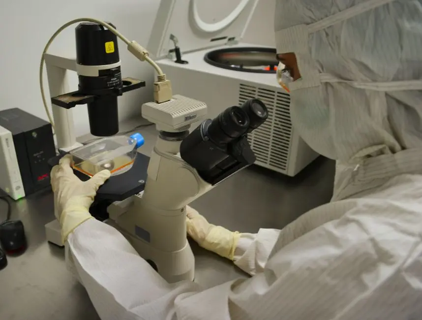
A sophisticated live-cell microscopy setup is more than just hardware; it is the intelligent integration of components orchestrated by powerful software. We utilize dedicated microscope control software to manage every aspect of the system: adjusting objectives, controlling illumination intensity and duration, operating the motorized stage, synchronizing environmental controllers, and precisely triggering the camera. This centralized control streamlines complex experiments and ensures reproducibility. The software must facilitate advanced image acquisition sequences, allowing us to program intricate protocols such as time-lapse imaging (capturing events over minutes to days), Z-stack acquisition (creating 3D volumetric data), and multi-channel imaging (observing multiple fluorescent markers simultaneously). Precision in timing and synchronization across these modes is critical for capturing dynamic biological interactions accurately.
Beyond acquisition, effective data handling and storage are paramount. Live-cell experiments generate vast amounts of data—terabytes are not uncommon for long-term, multi-channel runs. We must implement robust strategies for high-speed data transfer, secure storage (e.g., RAID arrays, network-attached storage), and efficient backup. Basic image processing capabilities within the acquisition software, such as real-time background subtraction or simple filtering, can aid in immediate quality control. However, for in-depth quantitative analysis, specialized post-acquisition analysis software (e.g., ImageJ/Fiji, commercial platforms like Imaris, Volocity) is indispensable. This software empowers us to track cell movement, measure intensity changes, quantify protein translocation, and perform colocalization analysis, transforming raw image data into profound biological insights. Building a workflow that seamlessly integrates acquisition with analysis unlocks the full narrative of cellular dynamics.
Optimizing Performance: Addressing Challenges and Integrating Advanced Techniques
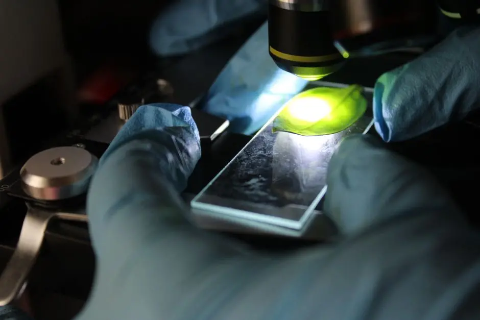
Even with state-of-the-art equipment, live-cell microscopy presents inherent challenges. Phototoxicity (light-induced damage to cells) and photobleaching (irreversible fading of fluorophores) are constant threats. We combat these by employing strategies such as using the lowest possible excitation light intensity, shortest exposure times, highly sensitive detectors (like sCMOS), and acquiring images only when biologically necessary. Environmental stress can also manifest subtly; ensuring our cells are accustomed to the imaging conditions before starting an experiment, using optimized media, and gentle handling techniques are crucial. Regular calibration of all environmental and optical components is a non-negotiable best practice to maintain peak system performance and data reliability. A proactive approach to troubleshooting focus drift, light source fluctuations, and software glitches ensures seamless operation.
To push the boundaries of discovery, we integrate advanced live-cell techniques that leverage and demand the robust equipment described. Techniques like FRAP (Fluorescence Recovery After Photobleaching) and FRET (Förster Resonance Energy Transfer) require precise laser control and synchronized detection for quantifying molecular dynamics and interactions. Optogenetics demands specific light sources for cell stimulation, often requiring synchronization with imaging acquisition. For resolving subcellular structures beyond the diffraction limit, live-cell super-resolution microscopy techniques (e.g., SIM, instant-STED) necessitate specialized optics, powerful lasers, and high-speed, high-sensitivity detection. Finally, the sheer volume and complexity of data generated require significant computational power—high-performance workstations with ample RAM and powerful GPUs are essential for efficient processing, rendering, and quantitative analysis. By mastering these challenges and integrating advanced tools, we transform live-cell microscopy into an unparalleled engine for biological exploration, revealing the exquisite ballet of life in real-time.
Key Takeaways
Microscope Platform & Optics
Prioritize inverted microscope systems with motorized stages for stability and flexibility. Essential optics include high Numerical Aperture (NA) objectives with long working distances (WD) and correction collars to ensure high resolution and adaptability to various sample conditions. Vibration isolation is crucial for artifact-free imaging.
Environmental Control
Robust temperature, CO2, and humidity control units are indispensable for maintaining physiological conditions. Integrate specialized specimen chambers and consider autofocus mechanisms to counteract focus drift over long experiments. Precise control prevents cell stress and ensures data integrity.
Illumination & Detection
Choose LED or laser light sources for precise, low-phototoxicity illumination. Select sCMOS cameras for a balance of sensitivity, speed, and field of view, or EMCCD cameras for ultra-low light applications. High-quality fluorescence filter cubes are vital for spectral purity.
Software & Data Management
Powerful microscope control software is key for orchestrating complex time-lapse, Z-stack, and multi-channel acquisitions. Implement robust data storage solutions and utilize specialized analysis software (e.g., ImageJ/Fiji) for quantitative insights. Ensure computational resources match data generation needs.
Optimization & Advanced Techniques
Actively mitigate phototoxicity and photobleaching by optimizing light dose and detector settings. Integrate specialized hardware and software for advanced techniques like FRAP, FRET, optogenetics, or super-resolution microscopy. Continuous calibration and a proactive approach to troubleshooting enhance experimental success and unlock deeper biological understanding.
FAQ
-
What is the most critical component for maintaining cell viability during long-term live-cell imaging?
The most critical component is a robust and precise environmental control system. This includes accurate temperature regulation (typically 37°C for mammalian cells), precise CO2 control to maintain media pH, and effective humidity control to prevent evaporation. Neglecting any of these factors can rapidly lead to cell stress, altered behavior, and even cell death, invalidating experimental results.
Advanced systems also incorporate autofocus mechanisms to counteract thermal drift, ensuring cells remain in focus over extended periods without additional stress from manual adjustments. -
How do I choose between an sCMOS and an EMCCD camera for live-cell imaging?
The choice hinges on your specific experimental needs, primarily regarding sensitivity, speed, and field of view.
sCMOS cameras offer an excellent balance, providing high sensitivity, very fast acquisition speeds, and a large field of view, making them suitable for most live-cell applications where you need to image dynamic events across many cells. They minimize phototoxicity due to their speed and efficiency.
EMCCD cameras, by contrast, offer unparalleled sensitivity, capable of detecting single photons. This makes them ideal for extremely low-light applications, such as imaging very dim fluorescent signals or single molecules, where minimizing light dose is paramount. However, they typically have a smaller field of view, can be more expensive, and may introduce read noise at lower EM gains. Consider your fluorophore brightness and the speed of the phenomena you wish to observe to make the optimal choice. -
What are common pitfalls in setting up and performing live-cell microscopy experiments?
Several common pitfalls can compromise live-cell microscopy experiments.
1. Poor environmental control: Inconsistent temperature, pH, or humidity leads to cell stress and altered physiology.
2. Phototoxicity and photobleaching: Excessive light exposure damages cells and fades fluorophores, necessitating careful optimization of light intensity, exposure time, and camera sensitivity.
3. Focus drift: Thermal changes can cause the sample to move out of focus; robust autofocus systems are crucial.
4. Vibrations: External vibrations (e.g., from building HVAC, nearby equipment) can blur images, requiring vibration isolation tables.
5. Insufficient data storage/processing: Live-cell experiments generate massive datasets, demanding high-capacity storage and powerful computational resources for analysis.
6. Suboptimal optics: Using objectives not corrected for temperature or media, or with low NA, compromises image quality.
Addressing these proactively through careful planning, proper equipment selection, and rigorous experimental protocols is vital for success.