
> Laboratory Techniques > Microscopy and Cellular Imaging > Unlocking Cellular Secrets: Confocal Microscopy Applications
Unlocking Cellular Secrets: Confocal Microscopy Applications
In the quest to decipher life's intricate mechanisms, vision is paramount. For centuries, biological research was constrained by the limits of conventional microscopy, offering mere glimpses into a 2D world. Today, the landscape has transformed. Confocal microscopy emerges as a revolutionary beacon, empowering scientists to penetrate deeper, visualize sharper, and analyze cellular and subcellular structures with unprecedented clarity. This advanced imaging technique has become an indispensable tool across diverse biological disciplines, from unraveling the mysteries of neurological pathways to charting the delicate dance of cellular organelles.
We stand at the precipice of discovery, ready to explore how this powerful technology is redefining our understanding of life itself. This deep dive will equip you with the strategic insights and practical knowledge necessary to harness its full potential, offering a specialized lens into the myriad applications that solidify confocal microscopy's role as a cornerstone in modern biological research. For those seeking to master advanced sophisticated techniques for biological structures, understanding confocal microscopy's capabilities is not merely an advantage; it is a fundamental requirement for groundbreaking scientific exploration.
Mastering the Confocal Advantage: Principles and Power
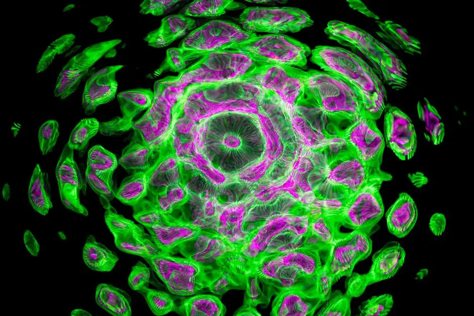
Confocal microscopy fundamentally transforms biological imaging by overcoming the inherent limitations of widefield microscopy. Its core distinction lies in the implementation of a pinhole aperture placed in the detection pathway. This ingenious design ensures that only light emitted from the focal plane reaches the detector, effectively rejecting out-of-focus fluorescence. This process, known as optical sectioning, allows for the acquisition of exceptionally sharp, thin optical slices through a specimen.
The power of confocal microscopy stems from its ability to construct stunning 3D reconstructions of complex biological structures, layer by layer, with unprecedented clarity. Key components orchestrate this precision: high-power lasers spanning various wavelengths excite specific fluorophores; precise scanning mirrors (galvanometer or resonant) systematically illuminate and collect light from each point in the sample; and sensitive detectors, often photomultiplier tubes (PMTs) or hybrid detectors, convert the photons into a digital signal. The result is an image with significantly improved signal-to-noise ratio and enhanced contrast, especially beneficial for thicker, more complex samples where conventional microscopy yields blurred, unusable data.
We must optimize critical parameters to fully harness this advantage. A common error involves improper pinhole setting: too small can reduce signal intensity unnecessarily, while too large compromises optical sectioning. Always calibrate the pinhole appropriately, typically to 1 Airy Unit, to balance resolution and signal. Furthermore, judiciously balancing laser power and detector gain is paramount to minimize photobleaching while maximizing the captured signal, preserving sample integrity during extended imaging. This strategic approach enables previously impossible visualizations of cellular architectures and tissue organization in their native 3D context, propelling our understanding of life's intricate designs.
Illuminating Cellular Dynamics: Applications in Cell Biology
Confocal microscopy is an indispensable tool for unraveling the intricate processes within individual cells, offering unparalleled insights into subcellular organization and dynamic events. One of its paramount applications lies in precisely determining the subcellular localization of proteins. Researchers routinely utilize fluorescent antibodies or genetically encoded reporters (such as GFP variants, mEmerald, mCherry) to tag specific proteins, visualizing their precise distribution within organelles, membranes, or cytoskeletal elements. Beyond mere visual identification, quantitative analysis, employing metrics like Pearson's or Manders' coefficients, provides robust data on molecular colocalization, indicating potential interactions or shared compartments.
The ability to perform live-cell imaging is another cornerstone application, allowing us to observe cellular dynamics in real-time. Confocal's reduced phototoxicity compared to widefield systems, especially when meticulously optimized, makes it ideal for tracking vesicle transport, mitochondrial fission/fusion, actin polymerization, or membrane trafficking over extended periods. Fast-scanning technologies, such as resonant scanners, minimize motion artifacts, preserving the fidelity of dynamic events. We must always employ rigorous controls: include single-label controls and unstained samples to accurately account for spectral bleed-through and intrinsic autofluorescence, ensuring the specificity and reliability of quantitative data. The acquisition of time-lapse sequences empowers us to extract critical kinetic information, transforming static snapshots into compelling narratives of cellular life.
For optimal results in live-cell imaging, maintaining a stable physiological environment is crucial. This involves temperature control, CO2 regulation, and appropriate culture media. Neglecting these environmental factors is a common pitfall that can compromise cell viability and experimental reproducibility. By meticulously managing both imaging parameters and cellular conditions, we unlock the full potential of confocal microscopy to reveal the dynamic ballet within living cells, pushing the frontiers of cell biological discovery.
Mapping Life's Blueprint: Developmental Biology and Neuroscience Insights
Shifting our focus to multicellular systems, confocal microscopy provides a critical lens for understanding complex processes in developmental biology and neuroscience. In developmental biology, it enables the visualization of embryonic development, organogenesis, and precise cell lineage tracing in intact embryos or organoids. Scientists track cell migration patterns, observe morphogenetic movements, and analyze gene expression within the context of whole, living organisms, revealing the intricate blueprints that guide development. The ability to generate high-resolution 3D images of these developing structures is paramount for deciphering how form and function emerge.
For neuroscience, confocal microscopy is transformative. It allows for the high-resolution mapping of neuronal circuits, visualization of dendritic spine dynamics associated with synaptic plasticity, observation of glial cell interactions, and assessment of blood-brain barrier integrity. When imaging deeper into tissue, conventional confocal might face limitations due to light scattering. To address this, two-photon microscopy stands out, employing longer-wavelength excitation light to reduce scattering and phototoxicity, making it ideal for in vivo brain imaging and penetrating several hundreds of micrometers into live tissue. This technique enables sustained, deep imaging without causing significant damage to the delicate neural environment.
Furthermore, to visualize structures within even thicker, opaque samples, tissue clearing techniques (e.g., CLARITY, CUBIC, iDISCO+) have revolutionized the field. When combined with confocal or two-photon microscopy, these methods render tissues transparent, allowing for unprecedented 3D visualization of entire organs or even whole organisms, from cellular resolution to macro-scale connectivity. A common error involves inadequate sample preparation for these thicker tissues, which can lead to light scattering and poor image quality. We advocate for rigorous adherence to established clearing protocols and the use of appropriate refractive index-matching mounting media. These strategic integrations enable us to decipher the complex wiring and dynamic events that govern biological development and brain function with unparalleled clarity.

Pushing the Boundaries: Advanced Techniques and Emerging Horizons
Confocal microscopy extends far beyond basic imaging, integrating advanced techniques that allow us to probe molecular function and interactions at high resolution. Fluorescence Recovery After Photobleaching (FRAP) is a powerful method to quantify molecular mobility within a cell. By photobleaching a specific region of interest with a high-intensity laser pulse and then monitoring the recovery of fluorescence as unbleached molecules diffuse back into the region, we derive crucial kinetic data on molecular diffusion rates and binding interactions. Similarly, Förster Resonance Energy Transfer (FRET) is employed to detect molecular interactions or conformational changes at sub-diffraction limited distances, typically <10 nm, providing direct evidence of protein-protein interactions or intramolecular dynamics. Precise FRET pair selection and rigorous control experiments are essential to avoid misinterpretation of results.
Further enhancing capabilities, techniques like photoactivation and photoconversion enable the tracking of specific cell populations or protein cohorts within dynamic systems. For example, photoactivatable GFP allows researchers to 'turn on' fluorescence in a subset of cells or proteins, then follow their fate or movement over time, offering profound insights into cellular lineage or protein trafficking. The integration of super-resolution microscopy variants (e.g., STED, SIM) with confocal platforms is breaking the diffraction barrier, revealing nanometer-scale details of cellular structures previously unattainable. While these are distinct techniques, many modern confocal systems offer modules or compatible setups to push resolution limits further.
The translational impact of confocal microscopy is also expanding rapidly. In clinical settings, it assists in pathology for rapid tumor margin assessment, diagnostics (e.g., corneal imaging for disease detection), and high-throughput drug screening in complex 3D cell cultures, offering a significant advantage over 2D models. Looking forward, the field is poised for further innovation with AI-driven image analysis, automating complex segmentation and quantitative measurements, and deeper tissue imaging through integration with techniques like light-sheet microscopy. We forge ahead, leveraging these advanced strategies to extract unprecedented biological information.
Optimizing Confocal Workflow: Best Practices and Troubleshooting
Achieving publishable quality confocal images requires meticulous attention to every step of the experimental workflow, from sample preparation to data analysis. Sample preparation is foundational: choosing the appropriate fixative (e.g., paraformaldehyde for proteins, methanol for microtubules) and optimizing fixation duration are critical for preserving cellular architecture and antigenicity. Rigorous immunostaining protocols, including proper blocking and washing, minimize non-specific binding. Selecting an appropriate mounting medium with refractive index matching and anti-fade reagents safeguards fluorophore integrity during imaging.
Fluorophore selection demands careful consideration of excitation/emission spectra to match available laser lines and filters, ensuring minimal spectral overlap during multiplexing. Prioritize photostable dyes for extended live-cell imaging to mitigate photobleaching. During image acquisition, we must strategically balance parameters: pixel dwell time influences signal-to-noise ratio and acquisition speed; line or frame averaging reduces random noise but increases acquisition time and photobleaching; and setting the Z-step size accurately, adhering to the Nyquist sampling theorem, is vital for high-fidelity 3D reconstructions. Over-saturating the detector by using excessively high laser power or gain is a common pitfall that destroys quantitative information and should be avoided by monitoring intensity histograms.
Post-acquisition, image processing and analysis are paramount. Deconvolution algorithms mathematically remove out-of-focus blur, enhancing resolution. 3D rendering software allows for sophisticated visualization. Quantitative tools for segmentation, intensity measurements, and colocalization analysis extract meaningful data. Common pitfalls include persistent autofluorescence (addressable with specific dyes, spectral unmixing, or tissue clearing) and chromatic aberration (mitigable with high-quality objectives and software correction). A systematic troubleshooting approach is key: isolate variables, re-evaluate protocols, and consult instrument specialists. Finally, implement robust data management practices with standardized naming conventions, secure archiving, and comprehensive metadata to ensure reproducibility and long-term data integrity. This optimized workflow is our blueprint for consistent, high-impact confocal research.
Key Takeaways
Core Confocal Advantages
Confocal microscopy revolutionizes biological imaging by employing a pinhole to eliminate out-of-focus light, delivering crisp optical sections. This fundamental principle enables superior 3D reconstruction, high signal-to-noise ratios, and reduced photobleaching, allowing precise visualization of cellular and subcellular structures within thick specimens.
Key Applications Across Biology
Confocal microscopy is indispensable across diverse biological fields. In cell biology, it facilitates detailed protein localization, live-cell dynamics, and quantitative co-localization studies. For developmental biology and neuroscience, it enables unprecedented visualization of embryogenesis, neuronal connectivity, and tissue organization, often leveraged with advanced techniques like two-photon imaging for deeper penetration.
Advanced Techniques & Future Outlook
Beyond basic imaging, confocal platforms support functional assays like FRAP (molecular mobility) and FRET (molecular interactions). Integration with super-resolution methods pushes resolution limits. The future involves deeper tissue imaging (e.g., light-sheet), AI-driven analysis, and expanding clinical applications, ensuring confocal microscopy remains at the forefront of biological discovery.
FAQ
-
What is the primary advantage of confocal microscopy over traditional widefield microscopy?
Confocal microscopy's primary advantage is its ability to perform optical sectioning. By using a pinhole to block out-of-focus light, it generates sharp, high-resolution images of specific focal planes within a thick sample, significantly reducing blur and enabling superior 3D reconstruction compared to the composite, blurred images from widefield microscopes. -
How does confocal microscopy achieve 3D imaging?
Confocal microscopy builds 3D images by acquiring a series of 2D optical sections at different depths (Z-stacks) within a specimen. Each section is free from out-of-focus blur. These individual sections are then computationally reconstructed to create a high-resolution, three-dimensional representation of the sample, allowing for detailed visualization of complex structures. -
What are common challenges in confocal microscopy, and how can they be mitigated?
Common challenges include photobleaching (loss of fluorescence signal), phototoxicity (damage to live cells), and autofluorescence (background signal from the sample itself). Mitigation strategies involve using lower laser power, faster acquisition speeds, optimizing fluorophore selection for photostability, using anti-fade mounting media, and applying tissue clearing techniques or spectral unmixing to reduce autofluorescence. -
Can confocal microscopy be used for live-cell imaging?
Absolutely. Confocal microscopy is widely used for live-cell imaging due to its reduced phototoxicity compared to widefield techniques (especially with optimized settings) and its ability to capture dynamic cellular processes in real-time. Resonant scanners allow for very fast image acquisition, minimizing motion artifacts and preserving cell viability during extended observations.