
> Laboratory Techniques > Microscopy and Cellular Imaging > Unlocking Cellular Secrets: Advanced Fluorescence Biomolecule Imaging
Unlocking Cellular Secrets: Advanced Fluorescence Biomolecule Imaging
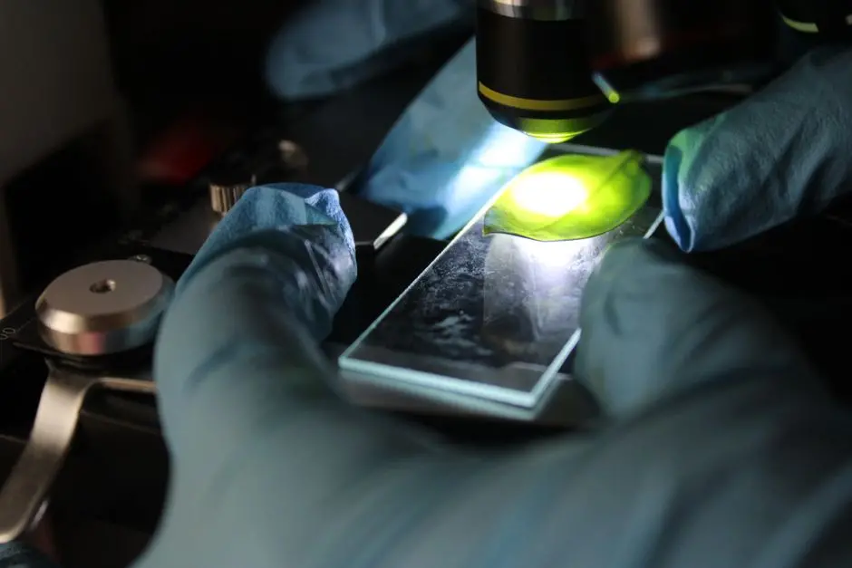
The biological realm, teeming with molecular interactions, largely remains an enigma without the power to witness its inner workings. How do proteins traffic within a cell? Where do specific enzymes catalyze their reactions? Unraveling these fundamental questions demands tools that transcend the limits of traditional microscopy. Fluorescence microscopy stands as a formidable vanguard in this quest, transforming the invisible into vibrant, actionable data. We embark on a journey to dissect the precise mechanisms by which this revolutionary technique illuminates biomolecules, providing unparalleled insights into cellular architecture and dynamic processes.
This comprehensive guide equips you with the strategic knowledge and tactical expertise to leverage fluorescence microscopy, turning complex biological phenomena into visually stunning and scientifically robust discoveries. Prepare to master the art and science of bringing cellular life into focus, integrating seamlessly with other advanced laboratory imaging methods to redefine our understanding of living systems. We forge ahead, transforming observational science into a dynamic exploration of life at its most fundamental level.
Illuminating the Invisible: The Core Mechanisms of Fluorescence Microscopy
Fluorescence microscopy operates on a principle of elegant simplicity yet profound capability: the specific emission of light by certain molecules, known as fluorophores, after absorbing light at a shorter wavelength. This fundamental process, termed fluorescence, is defined by the Stokes shift – the difference between the excitation and emission wavelengths. Unlike brightfield microscopy, which relies on light absorption and scattering to generate contrast, fluorescence imaging directly highlights target biomolecules, offering unmatched specificity and sensitivity within complex biological samples.
The architecture of a typical fluorescence microscope includes several critical components:
- Light Source: Emits a broad spectrum or specific wavelengths (e.g., LED, mercury lamp, laser).
- Excitation Filter: Selectively passes light at the fluorophore's excitation wavelength.
- Dichroic Mirror: Reflects excitation light towards the sample and transmits emitted fluorescent light towards the detector.
- Objective Lens: Focuses excitation light onto the sample and collects emitted fluorescent light.
- Emission Filter: Blocks residual excitation light, allowing only the longer-wavelength fluorescent emission to pass.
- Detector: Captures the emitted light signal (e.g., PMT, CCD, sCMOS camera).
We direct excitation light through the objective onto the sample. Fluorophores, bound to our biomolecules of interest, absorb this energy and subsequently emit light at a distinct, longer wavelength. This emitted light is then collected by the same objective, filtered through the dichroic mirror and emission filter, and finally recorded by the detector. This precise orchestration ensures that only the fluorescent signal from our target biomolecules contributes to the final image, providing a high signal-to-noise ratio crucial for discerning subtle biological events. We leverage this intricate interplay of light and matter to visualize the previously invisible.
Strategic Fluorophore Selection: Guiding Light to Specific Biomolecules
The success of fluorescence microscopy hinges on the judicious selection and application of fluorophores. These molecular beacons dictate the specificity, brightness, and longevity of our visualized biomolecules. We differentiate between several classes, each with unique advantages:
- Organic Dyes: Synthetic molecules offering high brightness and photostability (though some are prone to rapid photobleaching). Examples include DAPI for DNA staining, Phalloidin for actin, and the versatile Alexa Fluor or Cy dye series conjugated to antibodies. Their broad spectral range demands careful filter selection.
- Fluorescent Proteins (FPs): Genetically encoded such as GFP (Green Fluorescent Protein), RFP, and YFP. FPs are revolutionary for live-cell imaging because they allow direct fusion to a protein of interest, enabling visualization of its dynamics without cell fixation or permeabilization. This preserves physiological context.
- Quantum Dots (QDs): Semiconductor nanocrystals exhibiting exceptional brightness and photostability, with narrow emission spectra. However, potential toxicity and size can limit their utility in live-cell applications.
Labeling strategies are equally diverse:
- Immunofluorescence: A primary antibody specifically binds to the target biomolecule, followed by a secondary antibody conjugated with a fluorophore. This indirect method often amplifies the signal.
- Direct Labeling: The fluorophore is directly conjugated to a primary antibody or a small molecule probe, offering faster protocols but less signal amplification.
- Genetic Fusion: The gene encoding an FP is fused to the gene of interest, producing a chimeric protein that is inherently fluorescent.
- Bioorthogonal Chemistry: Methods like 'click chemistry' allow for highly specific labeling of engineered biomolecules in complex environments.
Best Practice: Always validate antibody specificity through controls and confirm fluorophore spectral compatibility with your microscope setup to avoid false positives or suboptimal signal. Common Error: Opting for fluorophores that photobleach too rapidly or exhibit significant spectral overlap with other labels or autofluorescence, compromising data integrity. We must ensure our choices serve the biological question with maximum clarity.
Unveiling Subcellular Detail: Advanced Fluorescence Modalities
While widefield fluorescence microscopy provides an excellent overview, its inherent limitation—out-of-focus blur from regions above and below the focal plane—obscures fine subcellular detail in thicker samples. To conquer this, we deploy advanced fluorescence modalities that redefine our spatial and temporal resolution, extracting deeper biological insights.
- Confocal Laser Scanning Microscopy (CLSM): This technique utilizes pinholes both before the detector and in the illumination path to physically block out-of-focus light. By scanning a focused laser beam across the sample and acquiring light from only a single optical plane, CLSM delivers crisp, high-resolution optical sections. We then reconstruct these sections to generate stunning 3D images of cellular structures, indispensable for visualizing biomolecules within complex, three-dimensional contexts like tissue sections or multicellular spheroids.
- Total Internal Reflection Fluorescence (TIRF) Microscopy: TIRF excels at imaging events occurring very close to the coverslip-sample interface, typically within <100 nm. It achieves this by generating an evanescent wave that only excites fluorophores in this narrow region. This dramatically reduces background fluorescence from the bulk of the cell, making it ideal for studying cell surface receptors, membrane dynamics, and initial adhesion events with exceptional signal-to-noise.
- Two-Photon Microscopy: This advanced method employs longer-wavelength, lower-energy photons (usually from a pulsed infrared laser) that excite fluorophores only when two photons simultaneously interact with the molecule. This non-linear excitation significantly reduces phototoxicity and scattering, allowing for deeper penetration into living tissues (up to several hundreds of micrometers) with minimal damage, crucial for *in vivo* imaging of biomolecules in intact organisms.
- Super-Resolution Microscopy (SRM): Pushing beyond the classical diffraction limit of light, SRM techniques like STED, PALM, and STORM achieve nanometer-scale resolution. While technically complex, these modalities enable us to visualize biomolecules and their organization with unprecedented detail, revealing structures and interactions previously unresolvable.
Insider Tip: Matching the microscopy technique to your specific biological question is paramount. For surface dynamics, TIRF offers superior signal-to-noise over confocal. For deep tissue imaging, two-photon microscopy is unmatched. We strategically select our tools to maximize discovery.
Mastering the Microscope: Best Practices and Troubleshooting for Optimal Imaging
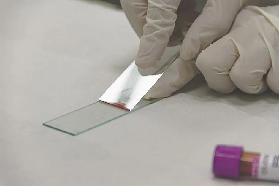
Achieving publication-quality fluorescence images requires meticulous attention to detail, from sample preparation to data interpretation. We systematically approach each stage to optimize our visualization of biomolecules.
- Sample Preparation is Foundational:
- Fixation: The choice of fixative (e.g., paraformaldehyde for protein preservation, methanol for microtubule structures) is crucial for maintaining cellular morphology while allowing probe access.
- Permeabilization: Detergents (e.g., Triton X-100) are essential to create pores in cell membranes, enabling antibodies and larger probes to access intracellular targets.
- Blocking: Using serum or BSA reduces non-specific binding of antibodies, minimizing background noise.
- Mounting Media: Select media that match the refractive index of the objective, preserve fluorescence (antifade reagents), and provide structural support.
- Mitigating Photobleaching and Autofluorescence:
- Photobleaching: The irreversible degradation of fluorophores upon light exposure. We combat this by minimizing excitation light intensity, reducing exposure times, using antifade agents, and sequencing acquisitions from most sensitive to most robust fluorophores.
- Autofluorescence: Background signal from intrinsic cellular components. Optimize emission filters, utilize spectral unmixing, and, in some cases, employ autofluorescence quenchers.
- Addressing Spectral Crosstalk: When fluorophores have overlapping emission spectra, their signals can bleed into each other's detection channels. We mitigate this through careful fluorophore selection (well-separated spectra), sequential acquisition of channels, and sophisticated spectral unmixing algorithms during image processing.
- Optimizing Data Acquisition Parameters: Precisely set detector gain, offset, pixel dwell time, and laser power to ensure optimal signal-to-noise without saturating the detector. Acquire sufficient Z-stacks for 3D reconstruction and time points for dynamic studies.
Common Pitfalls: Incorrect microscope settings, poor sample preparation leading to artifacts, and inadequate controls are pervasive issues. We rigorously include unstained, single-stained, and secondary-antibody-only controls to unequivocally identify true signals and background. Regular instrument calibration is non-negotiable. Actionable Advice: Always question your data and systematically troubleshoot any unexpected results. Master these practices, and we transform potential errors into opportunities for robust biological discovery.
Key Takeaways
Essence of Fluorescence: Key Takeaways for Biomolecule Visualization
Specificity & Sensitivity: Fluorescence microscopy utilizes targeted fluorophores to tag and visualize individual biomolecules with exceptional precision, overcoming the limitations of broad staining methods.
Fundamental Mechanism: The technique relies on fluorophores absorbing light at a specific wavelength (excitation) and emitting it at a longer wavelength (emission), enabled by a precisely configured optical system that filters light paths.
Diverse Labeling Strategies: Biomolecules are rendered fluorescent through various methods, including immunolabeling with antibodies, genetic encoding of fluorescent proteins, or direct chemical conjugation, each offering distinct advantages for fixed or live-cell studies.
Advanced Modalities: Techniques such as Confocal, TIRF, and Two-Photon microscopy significantly enhance resolution, imaging depth, or specificity. These advanced methods enable visualization of subcellular structures, dynamic surface events, or deep tissue layers, with super-resolution methods breaking the diffraction limit for nanoscale detail.
Operational Mastery: Successful fluorescence imaging demands meticulous sample preparation, careful fluorophore selection, effective mitigation of photobleaching and autofluorescence, and judicious data acquisition and analysis to derive robust, actionable biological insights. We champion this holistic approach for impactful science.
FAQ
-
What is the primary advantage of fluorescence microscopy over traditional brightfield microscopy?
Fluorescence microscopy offers unparalleled specificity and sensitivity, allowing researchers to visualize particular biomolecules within a complex cellular environment. Unlike brightfield, which relies on light absorption and scattering for contrast, fluorescence directly highlights target structures, often in living cells, providing dynamic insights that are simply impossible with conventional methods. We gain the power to precisely pinpoint and observe specific molecular events.
-
How do researchers choose the right fluorophore for their experiment?
Fluorophore selection is critical. We consider several factors: the nature of the target biomolecule, the experimental setup (live vs. fixed cells), required photostability, potential for spectral overlap with other labels, and the microscope's excitation and emission filter capabilities. Genetically encoded fluorescent proteins are ideal for live-cell dynamics due to their non-invasive nature, while bright organic dyes excel in fixed samples for high-resolution imaging. We match the tool to the task.
-
What are common challenges encountered in fluorescence microscopy and how can they be mitigated?
Common challenges include photobleaching (loss of fluorescence over time), autofluorescence (background signal from the sample), and spectral crosstalk (emission overlap between different fluorophores). We mitigate these by using antifade reagents, optimizing excitation light intensity and exposure, selecting fluorophores with well-separated spectra, employing spectral unmixing, and rigorously preparing samples to reduce background. Proactive strategy conquers these hurdles.
-
Can fluorescence microscopy visualize molecular interactions?
Absolutely. Advanced techniques like FRET (Förster Resonance Energy Transfer) and FLIM (Fluorescence Lifetime Imaging Microscopy) leverage the properties of fluorophores to detect interactions between biomolecules at nanometer distances. This allows us to map protein-protein interactions, conformational changes, and enzyme activities within living cells, providing profound mechanistic insights into dynamic cellular processes. We unlock the secrets of molecular partnership.