
> Laboratory Techniques > Microscopy and Cellular Imaging > Unraveling Cellular Dynamics: Light Microscopy's Core in Biology
Unraveling Cellular Dynamics: Light Microscopy's Core in Biology
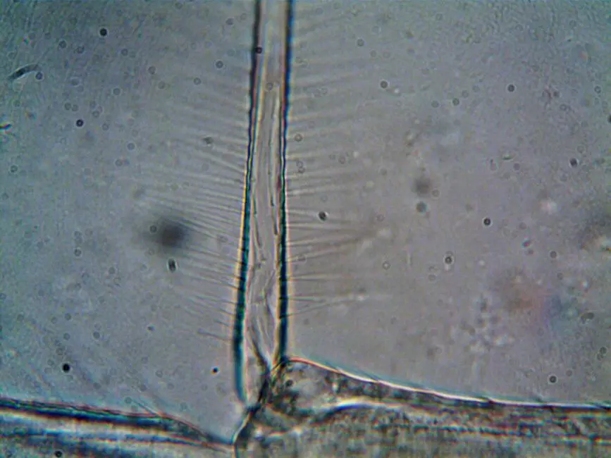
Imagine a world where the intricate dance of life remained hidden, an unseen ballet of cells and molecules. Before the advent of the microscope, our understanding of biology was limited to what the naked eye could perceive, leaving countless mysteries unsolved. Today, light microscopy stands as the bedrock of biological discovery, a relentless sentinel revealing the cellular architecture and dynamic processes that define existence. It is not merely a tool; it is a gateway to profound insights, a fundamental skill powering breakthroughs from disease diagnosis to genetic engineering.
This article will dissect the core principles and operational mechanisms that empower light microscopy, unveiling its indispensable role in modern biological research. We shall illuminate the physics of light interaction with specimens, meticulously detailing how magnification and resolution converge to render the invisible visible. Understanding these mechanics is crucial for every biologist seeking to extract meaningful data and push the frontiers of knowledge. Forge a deeper connection with the microscopic realm as we explore the essential capabilities that define effective laboratory imaging methods for biological structures. We unravel the science, empowering you to master this foundational technique and unlock new dimensions in your investigations.
Decoding Light's Embrace: Fundamental Principles of Microscopy
At its essence, light microscopy operates on the principle of harnessing photons to generate an enlarged image of a specimen. We initiate this process by directing a light beam through or onto a biological sample. The light then interacts with the specimen through various phenomena: absorption, reflection, refraction, and diffraction. These interactions modulate the light's amplitude, phase, and polarization, carrying vital information about the sample's physical properties. Our challenge lies in converting these subtle modulations into a discernible visual signal.
Two paramount concepts govern image quality: magnification and resolution. Magnification, the simple act of enlarging an image, is achieved through a series of lenses. However, mere enlargement is futile without the capacity to distinguish between two closely spaced points – this is where resolution becomes critical. The resolving power of a light microscope is fundamentally constrained by the Abbe diffraction limit, typically around 200-250 nanometers for visible light. This physical barrier dictates the smallest distance between two points that can be perceived as distinct, a limitation directly tied to the wavelength of light used and the numerical aperture (NA) of the objective lens. A higher NA, achieved by increasing the lens's light-gathering ability, directly translates to superior resolution, allowing us to discern finer details within the intricate landscape of a cell. We must continually push the boundaries of this physical constraint to reveal deeper biological truths.
The Microscope's Arsenal: Orchestrating Visual Discovery
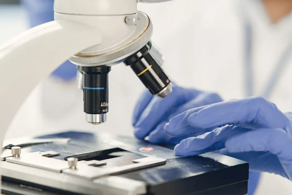
A light microscope is a meticulously engineered optical instrument, each component playing a critical role in the orchestration of image formation. We initiate the optical path with the light source, typically a halogen lamp or LED, providing stable and uniform illumination. This light travels through the condenser, a lens system positioned beneath the stage, which focuses the light beam onto the specimen. The condenser's numerical aperture (NA) adjustment is vital for controlling the angle of illumination and, consequently, the contrast and resolution of the image. Failure to optimize the condenser drastically compromises image quality, often resulting in glare or poor contrast.
The specimen itself rests on the stage, equipped with mechanical controls for precise X-Y movement. Above the specimen, the objective lens stands as the most critical component. This lens provides the primary magnification and, crucially, defines the microscope's resolution through its own numerical aperture. Objectives come in various magnifications (e.g., 4x, 10x, 40x, 100x) and optical corrections (e.g., achromat, plan-apochromat), each tailored for specific imaging tasks. Following the objective, the magnified intermediate image is further enlarged by the eyepiece (ocular), which projects the final image either to the observer's eye or to a camera sensor. Precise focusing is achieved through coarse and fine adjustment knobs, allowing us to navigate the specimen's depth and bring structures into sharp clarity. We must master the synergistic operation of these components to extract maximal information from our samples.
Expanding Vision: Pioneering Advanced Light Techniques
While brightfield microscopy provides a foundational view, its limitations for unstained, transparent biological samples are significant. We overcome these by employing advanced techniques that manipulate light's properties to enhance contrast and specificity. Phase contrast microscopy, for instance, ingeniously converts subtle phase shifts caused by variations in refractive index within a specimen into detectable brightness differences. This technique is invaluable for observing live, unstained cells and their dynamic processes, such as organelle movement or cell division, which would otherwise be invisible. We exploit the principle that light passing through different parts of a cell will be slightly delayed, creating phase differences that are then converted into amplitude differences by a phase plate.
Similarly, Differential Interference Contrast (DIC) microscopy utilizes polarized light and prisms to generate a pseudo-3D relief image. DIC excels at visualizing fine structures like mitochondria, vacuoles, or bacterial flagella, providing a striking sense of topography without the need for destructive staining. However, the true revolution in biological imaging emerged with fluorescence microscopy. This technique relies on fluorophores – molecules that absorb light at a specific excitation wavelength and emit light at a longer, characteristic emission wavelength. By attaching these fluorophores to specific cellular components (e.g., antibodies tagged with GFP or DAPI for DNA), we achieve unparalleled molecular specificity and sensitivity, allowing us to visualize particular proteins, organelles, or even genetic material against a dark, low-background field. Fluorescence microscopy has undeniably transformed our ability to localize and track molecules within living systems, propelling countless discoveries. We actively leverage these techniques to illuminate otherwise hidden aspects of cellular biology.
Mastering the Art: Optimizing Imaging & Avoiding Pitfalls
Achieving publication-quality images with light microscopy demands meticulous attention to detail and adherence to best practices. Our first critical step is impeccable sample preparation. This includes proper fixation to preserve cellular morphology, appropriate staining to enhance contrast, and correct mounting media with optimal refractive index. Crucially, the coverslip thickness must match the objective lens's specifications; deviation introduces spherical aberration, degrading resolution. A common pitfall for novices is neglecting Köhler illumination, a systematic alignment procedure that ensures uniform, bright illumination and optimal contrast, free from glare. We cannot overstate its importance: an incorrectly set Köhler illumination system sabotages image quality, irrespective of lens quality.
We must also become adept at identifying and mitigating common artifacts. Dust on lenses, air bubbles in mounting media, or smudges on slides can mimic cellular structures, leading to misinterpretation. Chromatic and spherical aberrations, inherent to lens optics, can also distort images; selecting high-quality, aberration-corrected objectives is a primary defense. Understanding the resolution limits of our chosen microscope prevents over-interpretation; we acknowledge that structures below the Abbe limit remain inaccessible to conventional light microscopy. Finally, establishing rigorous good practices—routine lens cleaning with appropriate solvents, proper microscope storage, and careful calibration of digital image acquisition parameters (exposure, gain)—safeguards the instrument's performance and the integrity of our data. We proactively identify and rectify these issues to ensure our images accurately reflect biological reality.
The Evolving Frontier: Charting Microscopy's Future
The journey of light microscopy is far from complete; we are actively charting new frontiers that push its capabilities beyond traditional limits. Live-cell imaging stands as a transformative domain, allowing us to observe dynamic biological processes in real-time, from cellular migration to protein trafficking. This demands precise environmental control (temperature, CO2) and the use of non-phototoxic fluorescent probes to maintain cell viability over extended periods. We leverage sophisticated time-lapse acquisition systems to capture these intricate temporal events, generating invaluable datasets on cellular kinetics.
The integration of automation and high-throughput screening is revolutionizing drug discovery and systems biology. Robotic stages, automated focusing, and intelligent software control entire imaging workflows, enabling us to screen thousands of samples efficiently and quantitatively. This generates massive datasets, underscoring the imperative for robust computational biology and advanced image analysis pipelines. Furthermore, Correlative Light and Electron Microscopy (CLEM) offers a powerful synergy, allowing us to pinpoint specific fluorescently labeled structures with light microscopy and then examine their ultrastructure at nanometer resolution using electron microscopy. This multi-scale approach provides unprecedented contextual understanding. While conventional light microscopy adheres to the Abbe limit, we acknowledge the emergence of super-resolution microscopy techniques (e.g., STED, PALM/STORM) that break this barrier, offering molecular-level resolution by exploiting advanced optical physics and computational reconstruction. These cutting-edge methods redefine what is visible, propelling biological research into an era of extraordinary detail and dynamic insight. We relentlessly innovate, pushing the boundaries of observation to uncover new biological principles.
Key Takeaways
Core Principles: Resolution and Magnification
Light microscopy's power hinges on its ability to magnify specimens and resolve fine details. Resolution, dictated by the numerical aperture (NA) of the objective and the wavelength of light, defines the smallest distance at which two points can be distinguished. Magnification simply enlarges the image. Both are crucial for revealing cellular structures, with resolution being the ultimate determinant of visual clarity and information content.
Essential Components: An Integrated System
A light microscope integrates a light source, condenser, stage, objective lens, and eyepiece. The light source illuminates the specimen, the condenser focuses this light, the objective provides primary magnification and resolution, and the eyepiece magnifies further for observation. Each component's precision, particularly the objective's NA, is vital for optimal image formation and data accuracy.
Advanced Techniques: Expanding Observational Capabilities
Beyond basic brightfield, techniques like phase contrast and Differential Interference Contrast (DIC) enhance visibility of unstained, live specimens by converting subtle phase shifts into detectable brightness variations. Fluorescence microscopy revolutionized cell biology by enabling specific labeling and visualization of molecules and structures through light excitation and emission, offering unparalleled specificity and sensitivity for molecular tracking and localization.
Operational Mastery: Best Practices and Pitfall Avoidance
Achieving high-quality results demands adherence to best practices, including proper Köhler illumination, meticulous sample preparation, and regular microscope maintenance. Recognizing and mitigating common artifacts (e.g., dust, aberrations) is crucial for data integrity. Understanding the physical limits of resolution, like the Abbe limit, prevents misinterpretation and guides appropriate technique selection, ensuring our scientific conclusions are robust.
FAQ
-
What is the most common mistake beginners make with light microscopes?
Neglecting Köhler illumination is a frequent oversight. It is paramount for achieving uniform illumination and maximizing resolution and contrast. Improper adjustment leads to uneven brightness, glare, and reduced image quality, obscuring fine details. Prioritize mastering this setup to ensure optimal data acquisition.
-
How critical is the numerical aperture (NA) in objective lenses for image quality?
The numerical aperture is the single most critical factor determining an objective lens's resolving power and brightness. A higher NA allows the objective to gather more diffracted light from the specimen, leading to superior resolution—the ability to distinguish between two closely spaced points—and a brighter image. Always prioritize higher NA objectives for detailed work and high-quality results.
-
Can conventional light microscopy truly visualize individual molecules?
Conventional light microscopy is fundamentally limited by the Abbe diffraction limit, typically around 200-250 nanometers. This means it cannot resolve structures smaller than this, including individual molecules or molecular complexes. While super-resolution techniques (like STED, PALM/STORM) have shattered this barrier, they operate on distinct principles beyond standard light microscopy, offering molecular-scale visualization.
-
What is the primary advantage of fluorescence microscopy over brightfield microscopy in biological research?
Fluorescence microscopy's primary advantage is its exceptional specificity and sensitivity. By using fluorescent probes that bind to specific cellular components, we can visualize particular molecules or structures within a complex cellular environment, often in living cells, with high contrast against a dark background. Brightfield, in contrast, relies on differences in light absorption and scattering, offering significantly less molecular specificity and often requiring staining that can alter cell viability.