
> Laboratory Techniques > Microscopy and Cellular Imaging > Master Electron Microscopy: TEM vs. SEM Dissected
Master Electron Microscopy: TEM vs. SEM Dissected
We stand at the precipice of biological discovery, where unraveling the intricate architecture of life demands tools of unparalleled precision. Electron microscopy, a cornerstone of modern biological research, empowers us to transcend the limitations of light and peer into the nanoscale world of cells, tissues, and microorganisms. Yet, within this powerful domain, two titans reign: Transmission Electron Microscopy (TEM) and Scanning Electron Microscopy (SEM). Each offers a unique window, but understanding their fundamental differences is not merely academic; it is a strategic imperative for any biologist aiming to extract maximum insight from their samples.
We will dissect the core principles, sample preparation intricacies, and distinct applications that define TEM and SEM, transforming your approach to cellular imaging. By the end of this deep dive, you will possess the clarity and expertise to confidently select the optimal technique for your specific research questions. Prepare to elevate your understanding of laboratory imaging methods for biological structures and unlock new dimensions in your biological investigations. Forge your path to precise observation and robust data generation.
The Core Principles: Unveiling Microscopic Worlds Through Electron Interaction
At the heart of both Transmission Electron Microscopy (TEM) and Scanning Electron Microscopy (SEM) lies the fundamental principle of utilizing electron beams instead of light to visualize samples, bypassing the diffraction limit inherent to light microscopy. However, their methods of electron-sample interaction fundamentally diverge, dictating the type of information each technique can yield. We recognize TEM as the technique for probing internal structures, while SEM excels at revealing surface topography.
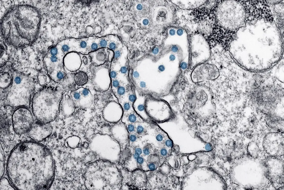
In TEM, a highly focused beam of electrons is transmitted through an ultrathin specimen. As electrons traverse the sample, they interact with its atomic structure. Denser regions scatter more electrons, while less dense areas allow more electrons to pass through. An electromagnetic lens system then collects these transmitted electrons, forming a two-dimensional projection of the sample's internal ultrastructure onto a detector. This process demands exceptionally thin samples, typically <100 nanometers, to ensure sufficient electron penetration. The resulting image provides astounding detail of organelles, macromolecular complexes, and cellular compartments, offering resolutions down to the sub-nanometer scale, often <0.2 nm under optimal conditions. This makes TEM indispensable for charting the physiological rigor of cellular machinery.
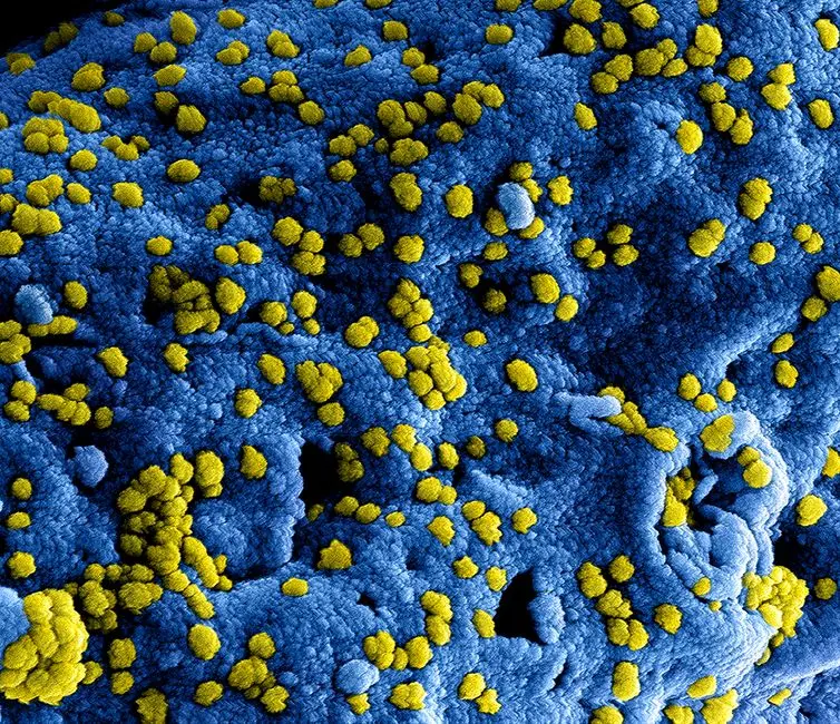
Conversely, SEM operates by scanning a finely focused electron beam across the surface of a sample. Instead of transmitting through, the primary electron beam interacts with the sample's surface atoms, generating various signals. The most commonly detected signals are secondary electrons (SEs), which are emitted from the very surface of the sample. The intensity of these emitted SEs varies with the surface topography: elevated areas emit more electrons than depressions. Backscattered electrons (BSEs) are another signal, originating from deeper within the sample and sensitive to atomic number. Detectors capture these electrons, and their signals are processed to construct a three-dimensional-like image of the sample's surface morphology. SEM typically achieves resolutions ranging from 1 to 20 nanometers, providing an excellent depth of field that conveys a strong sense of three-dimensionality. We wield SEM to explore the macroscopic architecture and intricate surface details, unlocking structural secrets that are hidden to TEM.
Preparing Your Sample: The Crucial Protocol Divergence for TEM and SEM
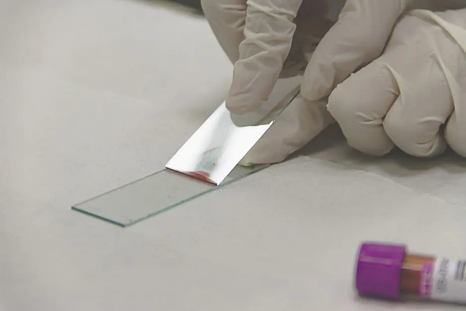
The quality and validity of electron microscopy data hinge critically on meticulous sample preparation. This stage is where TEM and SEM protocols diverge most significantly, each demanding specific approaches to preserve biological integrity, ensure electron interaction, and prevent artifact generation. We cannot overstate the importance of mastering these distinct methodologies, as errors here invariably compromise downstream analysis.
For TEM, the objective is to create an ultrathin, electron-transparent section that accurately represents the specimen's internal architecture. This typically involves a multi-step process: fixation, often using glutaraldehyde and osmium tetroxide, to chemically cross-link and stabilize cellular components; dehydration through increasing concentrations of ethanol or acetone to remove water; embedding in a rigid epoxy resin to support the fragile tissue; ultramicrotomy, where a diamond knife precisely cuts sections as thin as 50-100 nm; and finally, heavy metal staining (e.g., uranyl acetate and lead citrate) to enhance contrast by differential electron scattering. Common pitfalls include incomplete fixation leading to structural degradation, inadequate dehydration causing collapse during embedding, and sectioning artifacts such as chatter or compression. We relentlessly pursue perfection in these steps to guarantee accurate ultrastructural visualization.
SEM sample preparation, while still demanding, focuses on rendering the sample's surface conductive and stable under vacuum. The initial steps often mirror TEM: fixation (e.g., glutaraldehyde) to preserve morphology and dehydration. However, a critical divergence occurs with drying: biological samples must be dried without surface tension collapse. Critical point drying (CPD) is the gold standard, replacing a volatile solvent with liquid carbon dioxide, then gradually bringing it above its critical point, transforming it into a gas without forming a liquid-gas interface. This preserves delicate surface structures. The final, crucial step is sputter coating, where a thin layer (5-20 nm) of conductive material, typically gold or platinum, is deposited onto the sample surface. This prevents charge buildup from the electron beam, which would otherwise distort the image. Insider tip: an uneven or too-thick coat can obscure fine surface details. We strategize each preparation step to optimize surface integrity and conductivity, ensuring pristine topographic revelation.

Applications and Insights: Deploying TEM vs. SEM for Biological Questions
Selecting between TEM and SEM is not arbitrary; it is a strategic decision dictated by the specific biological question we aim to answer. Each technique carves its own niche, providing unique perspectives on the intricate world of biological systems. We must align the imaging modality with our research objectives to unlock maximal scientific value.
We deploy TEM when our mission is to unravel the ultrastructure of cellular organelles, analyze the intricate morphology of viruses, or precisely localize proteins within a cell. For instance, investigating mitochondrial cristae morphology, dissecting the internal organization of bacterial cells, or visualizing the budding process of enveloped viruses are quintessential TEM applications. Researchers also utilize TEM for immunogold labeling to pinpoint the exact subcellular location of specific proteins, providing invaluable insights into cellular function and pathology. In material science, TEM identifies nanoparticles' integration within biological matrices. The unparalleled resolution (<0.2 nm) and ability to generate 2D cross-sectional views make TEM the indispensable tool for delving deep into the hidden architecture that drives life processes. We leverage its power to discern the finest details of physiological structures.
Conversely, we activate SEM when our quest involves mapping the surface topography and three-dimensional arrangement of cells, tissues, or even entire organisms. SEM excels at revealing how cells interact with their extracellular matrix, examining the architecture of biofilms, or documenting changes in cell surface morphology during disease states or development. For example, visualizing the complex surface of a bacterial colony, mapping the cilia on a eukaryotic cell, or analyzing the intricate scales of an insect are ideal SEM tasks. Its superior depth of field (up to several millimeters) compared to TEM allows for a comprehensive, 3D-like appreciation of surface features, which is critical for understanding spatial relationships and macroscopic patterns. Resolutions typically range from 1 to 20 nm, providing sufficient detail for surface characterization. We utilize SEM to forge a visual understanding of surface interactions and environmental responses, transforming our perception of biological interfaces.

Operational Considerations and Future Trajectories: Maximizing Electron Microscopy Potential
Beyond the scientific distinctions, operational considerations significantly influence the choice and successful deployment of TEM and SEM in a research setting. We must navigate the practicalities of instrument operation, cost, and emerging technologies to optimize our biological investigations. Understanding these nuances empowers us to maximize the potential of electron microscopy.
Operational Complexity and Cost: TEM instruments generally demand a higher initial investment and possess greater operational complexity. Their maintenance requires specialized engineers, and user training is extensive, focusing on precise sample handling, vacuum systems, and intricate image acquisition parameters. SEMs, while still sophisticated, are often more accessible for routine surface imaging, with lower initial costs and a broader user base. Both require ultra-high vacuum environments, which necessitate rigorous maintenance and clean operating conditions. Consumables for TEM, such as high-purity embedding resins and diamond knives for ultramicrotomy, also contribute to ongoing operational expenses. We rigorously evaluate these factors to ensure sustainable and impactful research.
Limitations and Good Practices: Both techniques share common limitations, including the need for samples to withstand a vacuum, which historically precluded imaging of live biological specimens, and potential electron beam-induced radiation damage. We mitigate these through cryo-electron microscopy (Cryo-EM) for near-native state imaging. Good practices dictate regular instrument calibration, meticulous maintenance of vacuum systems, and a deep understanding of detector capabilities and limitations. We must also employ appropriate data analysis software, ensuring quantitative and unbiased interpretation of our high-resolution images. Insider tip: Always run control samples and maintain a detailed logbook for reproducibility.
Future Trajectories: The field of electron microscopy is dynamically evolving. Cryo-electron microscopy (Cryo-EM) revolutionizes structural biology by allowing us to image biological molecules and cellular structures in their near-native, hydrated states, without chemical fixation or staining. This technology earned the Nobel Prize in Chemistry in 2017. Furthermore, correlative light and electron microscopy (CLEM) integrates the functional insights from live-cell fluorescence microscopy with the ultrastructural detail of electron microscopy, forging a powerful combined approach. Advances in artificial intelligence (AI) and machine learning are poised to transform image acquisition, processing, and analysis, accelerating discovery and pushing the boundaries of what we can visualize. We champion these innovations to continuously enhance our understanding of biological complexity.
Key Takeaways
Core Distinctions: TEM vs. SEM
TEM (Transmission Electron Microscopy) probes internal ultrastructure at exceptionally high resolution (sub-nanometer), as electrons transmit through ultrathin sections. It provides 2D projection images of organelles and macromolecules.
SEM (Scanning Electron Microscopy) maps surface topography with a 3D-like appearance at nanometer-level resolution. Electrons scatter from the conductive-coated surface, revealing external features and spatial relationships.
Key Preparations Divergence
TEM demands intricate fixation, dehydration, resin embedding, ultrathin sectioning (<100 nm), and heavy metal staining for contrast. Focus: Electron transparency.
SEM involves fixation, dehydration, critical point drying (to prevent collapse), and conductive sputter coating (gold/platinum) to prevent charging. Focus: Surface stability and conductivity under vacuum.
Biological Applications & Optimal Use
Deploy TEM for questions regarding organelle ultrastructure, viral morphology, protein localization (immunogold labeling), and internal cellular pathology.
Utilize SEM for investigating cellular surface features, biofilm architecture, tissue morphology changes, and overall 3D surface topography, benefiting from its superior depth of field.
Operational Dynamics & Future Outlook
Operational: TEM requires higher investment and specialized expertise; SEM is generally more accessible. Both demand high vacuum, leading to challenges with live samples and potential radiation damage.
Future: Cryo-EM is revolutionizing structural biology by imaging near-native states. Correlative Light and Electron Microscopy (CLEM) integrates functional and structural insights. AI is enhancing image analysis, pushing the boundaries of biological visualization.
FAQ
-
Which provides higher resolution, TEM or SEM, and why?
Transmission Electron Microscopy (TEM) typically provides significantly higher resolution, often achieving sub-nanometer detail (e.g., <0.2 nm) compared to Scanning Electron Microscopy (SEM) which generally ranges from 1 to 20 nm. TEM's superior resolution stems from the fact that electrons pass through the sample, allowing for imaging of internal structures at very high magnification, where the wavelength of electrons is much smaller than that of light.
-
Can live biological samples be viewed using TEM or SEM?
Traditionally, no, due to the high vacuum required by both TEM and SEM, which desiccates and damages live samples. However, advancements like cryo-electron microscopy (Cryo-EM) allow for imaging of samples in a frozen, near-native, hydrated state, effectively circumventing the vacuum limitation for biological structures. Environmental SEM (ESEM) can also accommodate samples under partial vacuum, allowing some hydrated samples to be imaged, though usually at lower resolution.
-
What is the main difference in the type of image produced by TEM versus SEM?
TEM produces a two-dimensional, projection image revealing the internal ultrastructure of the sample, akin to an X-ray of a cellular cross-section. SEM, on the other hand, generates a three-dimensional-like image showcasing the surface topography and morphology of the sample. This difference dictates their primary applications: TEM for internal cellular architecture, SEM for external surface features.
-
Why is sample preparation considered the most critical stage for electron microscopy?
Sample preparation is paramount because electron beams operate in a vacuum and can easily damage or interact poorly with untreated biological tissues. Proper preparation (fixation, dehydration, embedding, sectioning/coating) preserves the sample's native structure, prevents artifacts, ensures stability under vacuum and electron beam exposure, and allows for effective electron interaction (transmission or scattering) to generate a high-quality, representative image. Errors in this stage directly compromise the scientific validity of the resulting data.