
> Laboratory Techniques > Microscopy and Cellular Imaging > Illuminate Molecular Architectures: Electron Microscopy in Biology
Illuminate Molecular Architectures: Electron Microscopy in Biology
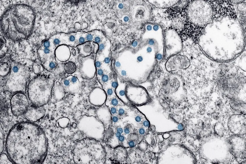
In the relentless pursuit of understanding life’s fundamental processes, our vision is often limited by the tools at hand. While light microscopy reveals the vibrant dance of living cells, its inherent resolution limits obscure the intricate molecular machinery driving these phenomena. To truly decipher the secrets held within the smallest cellular components—the proteins, nucleic acids, and macromolecular complexes—we must plunge deeper.
This article embarks on an expedition into the microscopic realm, showcasing how electron microscopy (EM) transcends these limitations, providing unparalleled detail critical for molecular biology. We will explore the transformative applications that empower researchers to visualize structures at atomic resolution, fundamentally reshaping our comprehension of biological function and disease mechanisms. Prepare to discover the essential role EM plays among advanced laboratory imaging methods for biological structures, propelling breakthroughs in structural biology and beyond. Forge understanding with us as we unveil the power of electron microscopy to unlock molecular mysteries.
Mastering Electron Microscopy Foundations for Molecular Insights
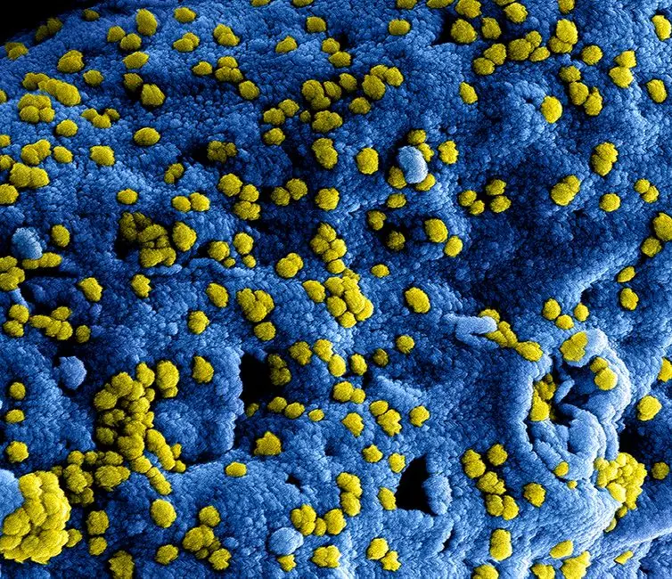
To truly conquer the complexities of molecular biology, we must first master the foundational principles that grant us access to the nanoscale. Electron microscopy, particularly Transmission Electron Microscopy (TEM) and Scanning Electron Microscopy (SEM), operates on a principle fundamentally distinct from light microscopy: it utilizes a beam of electrons instead of photons. This critical shift allows for significantly shorter wavelengths, directly translating into superior resolution, often reaching atomic levels. While SEM excels in providing detailed surface topography with striking three-dimensional perspectives, it is TEM that unveils the intricate internal architecture of biological specimens, a cornerstone for molecular investigations, especially concerning large macromolecular complexes. High-brightness electron sources, such as field emission guns, are paramount here, delivering coherent electron beams essential for high-resolution imaging.
The journey from a biological sample to a high-resolution electron micrograph is a meticulous one, demanding precision at every stage. Traditional TEM methods involve intricate sample preparation: chemical fixation to preserve native structures, dehydration, embedding in resin, and ultra-thin sectioning (typically 50-100 nm). Contrast is then introduced through heavy metal staining, which differentially scatters electrons, generating the image. However, a revolution has swept through the field with the advent of cryo-electron microscopy (cryo-EM). This groundbreaking technique involves flash-freezing samples, typically on specialized EM grids, at extremely low temperatures, vitrifying them in a thin layer of amorphous ice. This crucial step eliminates the need for harsh chemical fixation and staining, preserving molecules in their near-native, hydrated state. We bypass artifacts induced by traditional methods, capturing transient conformational states that are vital for understanding dynamic biological processes, a common error in older approaches. Careful blotting of the grid is an insider tip for achieving optimally thin ice layers, critical for high-resolution.
Understanding these distinctions is paramount. Cryo-EM has fundamentally redefined our approach, allowing us to image individual macromolecules, viruses, and even entire organelles with unprecedented clarity. The resolution achieved pushes the boundaries of visualization, allowing for the direct observation of protein folds and molecular interactions. We are no longer limited to inferring structure; we directly observe it. This expertise in choosing the correct EM technique and mastering sample preparation—including understanding electron dose limitations to minimize radiation damage—is the bedrock upon which all subsequent molecular insights are built. We unlock new dimensions of biological understanding, moving from macroscopic observations to the precise atomic arrangements governing life itself, forging a path towards complete molecular blueprints.
Dissecting Molecular Machines: Protein Structure Determination
The accurate determination of protein structure is not merely an academic exercise; it is the linchpin for understanding function, designing targeted therapies, and reverse-engineering biological processes. Electron microscopy, particularly advanced cryo-EM methodologies, has become an indispensable powerhouse in this arena. We are now able to dissect the intricate architectures of even the most challenging molecular machines, overcoming previous limitations imposed by crystallography for large, flexible complexes or NMR spectroscopy for molecules exceeding ~50 kDa. The ability to resolve protein heterogeneity and multiple conformational states within a single sample is a distinct advantage.
Single-particle analysis (SPA) stands at the forefront of this revolution. This technique involves imaging millions of identical copies of a macromolecule, each captured in a slightly different orientation, within a thin layer of vitreous ice. The computational pipeline for SPA is sophisticated: it begins with motion correction to compensate for sample movement during electron exposure, followed by contrast transfer function (CTF) estimation to correct for optical aberrations, and automated particle picking to identify individual molecules. Next, 2D and 3D classification steps sort particles by orientation and conformational state, enabling averaging to reconstruct a high-resolution three-dimensional map. This iterative refinement process often achieves near-atomic or even atomic resolution, allowing for the visualization of individual amino acid side chains. Examples abound: we have meticulously mapped the structure of the ribosome, the intricate protein synthesis factory (e.g., at 1.5 Å resolution), revealing its complex catalytic mechanisms; elucidated the architectures of diverse viral capsids, critical for vaccine development; and deciphered the structures of membrane proteins, notoriously difficult to crystallize, which are central to cellular signaling and major drug targets (e.g., GPCRs). A common pitfall here is insufficient particle numbers or misclassification, leading to blurred maps.
Beyond isolated macromolecules, cryo-electron tomography (cryo-ET) extends EM’s power to visualize molecular machines directly within their native cellular context. By tilting a vitrified sample through a range of angles and acquiring a series of 2D images, we generate a 3D tomogram, a volumetric reconstruction of a cellular region. Subtomogram averaging then allows us to identify and average multiple instances of the same macromolecular complex within this crowded environment, revealing their in situ structures and interactions, providing unparalleled contextual insights. This method has illuminated the architecture of cytoskeletal filaments, revealed the arrangement of nuclear pore complexes, and visualized bacterial flagellar motors within living cells. The impact on drug discovery is profound; understanding the precise binding sites and conformational changes of target proteins at an atomic level in their native environment accelerates rational drug design, allowing us to forge more effective and specific therapeutic agents. We are transforming drug development from a trial-and-error process into a targeted, structure-driven endeavor, directly leveraging molecular blueprints.
Charting the Cellular Landscape: Ultrastructure and Organelle Dynamics
Understanding life demands more than isolated molecular structures; we must comprehend how these molecules assemble and interact within the dynamic, crowded environment of a cell. Electron microscopy offers a unique vantage point into this cellular landscape, providing unparalleled detail of ultrastructure and allowing us to chart the intricate dance of organelles and their interactions. For decades, conventional EM has been the gold standard for visualizing cellular architecture, revealing the complex folding of the endoplasmic reticulum, the precise organization of mitochondrial cristae, and the intricate cytoskeletal networks that define cell shape and motility. These visualizations are not static snapshots; they unveil the very blueprints of cellular life, fundamental for understanding normal physiology and pathological states. We frequently leverage quantitative EM approaches, such as stereology, to derive robust statistical data on organelle volumes, membrane surface areas, and particle densities, moving beyond qualitative observation to objective measurements.
The power of EM extends beyond simple morphology. By meticulously preparing tissues and employing advanced 3D reconstruction techniques, such as serial sectioning TEM or serial block-face SEM, we can map organelle-organelle contacts and understand the precise spatial relationships that govern cellular processes. For instance, EM reveals the close apposition of mitochondria and ER in areas of lipid synthesis or calcium signaling—forming "Mitochondria-Associated Membranes" (MAMs)—shedding light on their functional interplay. Similarly, we can observe the critical connections between the ER and peroxisomes in lipid metabolism. This detailed contextual information is vital for unraveling mechanisms of disease, such as neurodegeneration where mitochondrial dysfunction and ER stress are key players, or understanding infectious diseases where pathogens hijack host cell machinery. We directly observe the pathological reorganization of cellular compartments, providing crucial clues for therapeutic intervention and pinpointing common errors in cellular homeostasis.
A particularly potent strategy in this pursuit is Correlative Light and Electron Microscopy (CLEM). This innovative approach combines the benefits of both worlds: the broad overview, live-cell imaging capabilities, and molecular specificity (via fluorescent tags) of light microscopy, with the ultra-high resolution and ultrastructural detail of electron microscopy. Researchers first identify specific events or tagged molecules in living cells using fluorescence microscopy, then precisely locate and image those exact regions at high resolution using EM after sample preparation. This powerful synergy allows us to localize specific proteins or processes within the rich ultrastructural context of the cell. For example, tracking a vesicle budding event in live cells via fluorescence, then performing CLEM to visualize the precise membrane curvature and protein machinery involved at an ultrastructural level, provides an unprecedented mechanistic understanding. Challenges include accurate registration of LM and EM images and maintaining sample integrity during transfer, but best practices involve fiducial markers. We are forging a bridge between dynamic molecular events and their tangible structural reality, accelerating discoveries in cell biology and disease pathogenesis, and delivering a comprehensive view of cellular function in situ.
The Next Horizon: Advanced EM Technologies and Challenges
The frontier of electron microscopy is in constant flux, driven by relentless innovation that pushes the boundaries of resolution, speed, and analytical power. We are witnessing a golden age where technological advancements are rapidly transforming what was once considered impossible. One transformative technique is Focused Ion Beam-Scanning Electron Microscopy (FIB-SEM). This powerful hybrid instrument allows for serial sectioning of a sample, literally milling away nanometer-thick layers using a focused ion beam, followed by high-resolution imaging of each freshly exposed surface with an SEM. By acquiring hundreds or even thousands of these images, we can reconstruct large, complex volumes of cells or tissues in 3D with isotropic resolution (e.g., typically 5-10 nm), providing an unprecedented view of interconnected cellular networks, organelle morphology, and synaptic connections in their native context. This volumetric imaging capability is particularly crucial for mapping neural circuits, understanding tissue architecture in developmental biology, or characterizing viral infection patterns throughout entire cells. A critical best practice is precise alignment of the FIB and SEM beams to minimize artifacts.
Beyond imaging, the development of direct electron detectors (DEDs) has revolutionized cryo-EM. Unlike traditional detectors, DEDs directly detect electrons without scintillation, offering significantly higher sensitivity and faster readout speeds. This enables movies of electron exposures, allowing for computational correction of beam-induced sample motion and resulting in dramatic improvements in signal-to-noise ratio and resolution. This technological leap is a primary driver behind the "resolution revolution" in cryo-EM, making near-atomic resolution commonplace for many protein complexes, with resolutions now routinely reaching 2-3 Å or even better for stable complexes. Coupled with these hardware advancements, sophisticated computational methods and artificial intelligence (AI) are now indispensable. Machine learning algorithms optimize image processing, particle picking, classification, and 3D reconstruction, dramatically accelerating data analysis and enhancing the quality of structural models. We leverage AI to identify subtle features, correct for imperfections, and extract maximum information from inherently noisy data, significantly reducing manual intervention and processing time, which were common bottlenecks in the past.
However, navigating these frontiers presents its own set of challenges. Sample preparation, while advanced (e.g., cryo-FIB milling for thinning thicker samples for cryo-ET), remains a bottleneck for many biological systems, particularly for large, heterogeneous tissues or dynamic processes that are difficult to vitrify perfectly. Radiation damage from the electron beam is an inherent limitation for biological samples, requiring careful dose management and cryogenic temperatures. Interpreting complex 3D data volumes, especially from techniques like FIB-SEM or cryo-ET, demands specialized bioinformatics and visualization expertise. Despite these hurdles, the trajectory of EM development is clear: towards even higher resolution, faster data acquisition, and increasingly sophisticated integration with other imaging modalities, such as mass spectrometry imaging. We envision a future where EM routinely visualizes molecular interactions in situ with atomic precision, offering a complete picture of life’s machinery from the atomic scale to the entire organism. We are not just observing; we are decoding the very language of biology, piece by intricate piece, to forge a new era of understanding.
Key Takeaways
Core EM Strengths
Electron microscopy (EM) transcends the resolution limits of light microscopy by using electron beams, enabling visualization of molecular and cellular structures at atomic to nanoscale levels. This foundational advantage is crucial for molecular biology, where understanding the intricate details of proteins, organelles, and their interactions is paramount for deciphering life's processes and disease mechanisms.
Key EM Techniques
Two principal EM techniques dominate: Transmission Electron Microscopy (TEM) for internal structures and Scanning Electron Microscopy (SEM) for surface topography. Crucially, cryo-electron microscopy (cryo-EM) has revolutionized the field by imaging biological samples in their near-native state, avoiding fixation artifacts and enabling high-resolution structural determination of delicate molecular machines like proteins and viral capsids.
Impact on Molecular Biology
EM is indispensable for: Protein Structure Determination (via single-particle analysis for purified complexes and cryo-electron tomography for in-situ visualization within cells), and Cellular Ultrastructure Analysis (mapping organelle architecture, interactions, and understanding disease mechanisms at a subcellular level). It significantly empowers rational drug discovery by revealing precise molecular targets and their interactions.
Future Outlook and Challenges
Advanced EM technologies like Focused Ion Beam-Scanning Electron Microscopy (FIB-SEM) for volumetric imaging, direct electron detectors (DEDs) for enhanced resolution, and AI-driven computational methods continue to push boundaries. Despite challenges like meticulous sample preparation and inherent radiation damage, EM is poised to offer even more comprehensive and dynamic insights into biological systems at an unprecedented level of detail, truly decoding life's molecular language.
FAQ
-
What are the primary advantages of Electron Microscopy over light microscopy in molecular biology?
Electron Microscopy (EM) offers significantly higher resolution, allowing visualization of structures at the nanoscale, down to individual atoms. This capability is critical for molecular biology, where we need to resolve proteins, nucleic acids, and macromolecular complexes, which are invisible to light microscopy due to its resolution limit imposed by the wavelength of light. It provides direct structural insights unavailable through light-based methods.
-
What is cryo-EM and why is it revolutionary for structural biology?
Cryo-electron microscopy (cryo-EM) is a technique where biological samples are rapidly frozen in a thin layer of vitreous ice, preserving them in a near-native, hydrated state without the need for chemical fixation or staining. It is revolutionary because it enables the determination of protein and complex macromolecular structures at atomic or near-atomic resolution, especially for samples that are difficult to crystallize, providing unprecedented insights into their function and dynamics directly from solution.
-
What are the main challenges in applying EM to molecular samples?
Key challenges include meticulous sample preparation (e.g., achieving thin, vitreous ice for cryo-EM), radiation damage to delicate biological samples from the electron beam, which necessitates low-dose imaging, and the intensive computational processing required to reconstruct high-resolution 3D models from noisy 2D projections. Additionally, interpreting complex volumetric data demands specialized bioinformatics and visualization expertise, and sample heterogeneity can complicate analysis.
-
How does EM contribute to drug discovery?
EM plays a crucial role in drug discovery by providing high-resolution structural information of target proteins, including membrane proteins and large protein complexes, which are often intractable by other methods. Understanding the precise 3D architecture of these targets and how potential drug candidates interact with them at an atomic level enables rational drug design, accelerating the development of more specific and effective therapeutic agents. It helps validate target binding sites and mechanisms of action.