
> Laboratory Techniques > Microscopy and Cellular Imaging > Conquering Live-Cell Imaging: Navigating Key Experimental Hurdles
Conquering Live-Cell Imaging: Navigating Key Experimental Hurdles
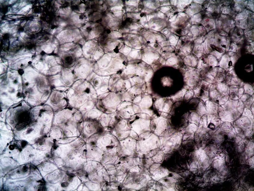
The dynamic world within living cells reveals secrets inaccessible through static observation. Live-cell imaging, a cornerstone of modern biology, unveils these processes in real-time, providing unparalleled insights into cellular function, disease progression, and therapeutic responses. However, capturing these transient moments demands overcoming a formidable array of technical and biological challenges. We empower researchers to master these complexities, transforming potential pitfalls into pathways for discovery.
This definitive article dissects the core hurdles – from maintaining physiological conditions to mitigating phototoxicity and ensuring long-term cellular viability. We architect a strategic framework for success, guiding you through best practices and cutting-edge solutions. Unlock the full potential of your experiments, moving beyond mere observation to definitive biological breakthroughs, leveraging advanced laboratory imaging methods for biological structures.
Mastering the Microenvironment: Preserving Physiological Conditions and Cell Viability
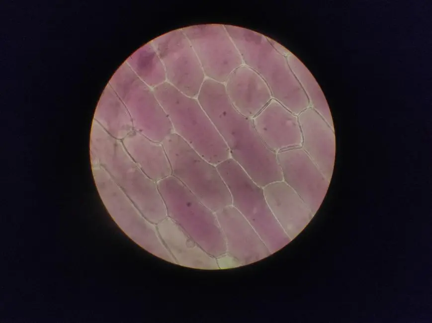
Maintaining a stable, healthy cellular environment throughout extended imaging sessions represents a foundational challenge in live-cell microscopy. Any deviation from optimal physiological parameters compromises cell viability, alters natural biological processes, and introduces experimental artifacts, rendering results unreliable. We must orchestrate precise control over temperature, CO2/pH, osmolarity, and sterility to ensure our observations genuinely reflect cellular behavior.
Key considerations include:
- Temperature Control: Cells thrive within a narrow temperature range (typically 37°C for mammalian cells). Rapid temperature fluctuations induce stress, alter metabolic rates, and impact protein function. Implement a robust environmental chamber around the microscope, ensuring uniform temperature distribution across the sample. Calibrate regularly and monitor using external sensors.
- CO2 and pH Regulation: Most cell culture media are buffered with bicarbonate, requiring precise CO2 levels (typically 5%) to maintain physiological pH (7.2-7.4). Fluctuations lead to acidosis or alkalosis, severely impacting cellular processes. Utilize dedicated CO2 incubators integrated with the imaging system, capable of precise gas mixing and humidity control.
- Osmolarity and Media Evaporation: Prolonged imaging can lead to media evaporation, increasing osmolarity and stressing cells. Employ specialized imaging dishes with low evaporation rates, add mineral oil overlays, or use microfluidic devices for continuous media perfusion. Ensure culture media contain appropriate supplements and serum, refreshed if imaging extends beyond 24 hours.
- Sterility: Contamination by bacteria, fungi, or mycoplasma jeopardizes entire experiments. Execute rigorous aseptic techniques during sample preparation and handling. Utilize sterile culture vessels and media, and operate within a laminar flow hood. Implement regular cleaning and sterilization protocols for all equipment in contact with samples.
Failing to address these parameters constitutes a classic error, leading to cellular distress and spurious data. Our strategy focuses on proactive environmental control, establishing an unshakeable foundation for reliable live-cell observations.

Combatting Phototoxicity and Photobleaching: Safeguarding Dynamic Processes
Illuminating living cells, particularly with high-intensity light sources or extended exposure, inflicts inevitable damage. This phototoxicity manifests as cellular stress, DNA damage, altered signaling pathways, and ultimately, cell death, corrupting the very dynamics we aim to capture. Concurrently, fluorophores degrade over time, leading to photobleaching and a loss of signal, severely limiting observation duration. Overcoming these twin challenges is paramount for high-fidelity live-cell imaging.
Strategic approaches involve:
- Minimize Light Exposure: This is the golden rule. Employ the lowest possible laser power or lamp intensity that yields a detectable signal. Shorten exposure times per frame and increase the interval between frames (lower frame rate) when fast dynamics are not strictly necessary. Use neutral density filters to finely tune light intensity.
- Choose Robust Fluorophores: Select fluorophores with high photostability and brightness. Genetically encoded fluorescent proteins (e.g., mEGFP, mCherry) designed for live-cell imaging often outperform synthetic dyes in terms of photostability and reduced toxicity. Avoid fluorophores prone to reactive oxygen species (ROS) generation.
- Optimize Imaging Modes: Explore alternative imaging techniques. Light-sheet microscopy (SPIM) illuminates only the focal plane, drastically reducing phototoxicity by orders of magnitude compared to widefield or confocal. Spinning disk confocal microscopy offers faster acquisition with lower light doses than point-scanning confocal. Total Internal Reflection Fluorescence (TIRF) microscopy restricts excitation to the basal membrane, ideal for surface events.
- Employ Antioxidants and ROS Scavengers: In some cases, adding antioxidants (e.g., N-acetylcysteine, Trolox) to the imaging medium can help mitigate ROS-induced damage, though this must be carefully validated for specific cell types and experiments to avoid unintended biological effects.
- Advanced Detection Systems: Utilize highly sensitive detectors like sCMOS or EMCCD cameras, which allow for lower excitation intensities while maintaining sufficient signal-to-noise ratios.
We forge a proactive approach to light management, balancing the need for clear signals with the imperative to preserve cellular health. This careful calibration ensures observed dynamics are intrinsic, not artifacts of our imaging strategy.
Pushing Boundaries: Achieving High Spatial, Temporal, and Z-Resolution
Live-cell imaging demands a delicate balance between observing fine structural details (spatial resolution), capturing rapid events (temporal resolution), and resolving structures through cellular depth (Z-resolution). Often, optimizing one comes at the expense of another, creating a critical challenge in experimental design. We must strategically select instrumentation and imaging parameters to achieve the resolution necessary for our specific biological questions without introducing excessive light dose or compromising cell viability.
Strategic solutions involve:
- Objective Lens Selection: Utilize high numerical aperture (NA) objectives (e.g., 1.3 NA to 1.49 NA oil immersion or water immersion) for superior spatial resolution and light collection. Matched refractive index immersion oils are crucial. For deep tissue imaging, specialized long working distance objectives are essential.
- Detector Optimization: Fast and sensitive detectors like sCMOS cameras (for widefield/light-sheet) or resonant scanners (for confocal) enable higher frame rates for temporal resolution. Smaller pixel sizes on detectors, when paired with appropriate magnification, contribute to spatial resolution.
- Focus Stability: Drift, caused by thermal expansion or mechanical instability, plagues long-term imaging, leading to loss of focus and blurred images. Implement active focus stabilization systems (e.g., hardware autofocus modules) that continuously monitor and correct for focus changes. Ensure the microscope and environmental chamber are thermally equilibrated before starting experiments.
- Z-Resolution and Optical Sectioning: For 3D imaging, optical sectioning techniques are vital. Confocal microscopy provides excellent Z-resolution by rejecting out-of-focus light. Light-sheet microscopy excels at rapid 3D imaging with minimal phototoxicity. Deconvolution algorithms can computationally enhance Z-resolution and improve signal-to-noise.
- Balancing Trade-offs: High temporal resolution often requires increased light exposure and lower spatial sampling. Conversely, achieving super-resolution pushes the limits of light dose and acquisition speed. We must precisely define the biological question to prioritize resolution parameters and accept necessary trade-offs. For instance, single-molecule tracking demands high temporal and spatial resolution, often at the cost of observing only a small field of view.
We architect precise control over optical parameters, ensuring our experimental setup is finely tuned to the biological question. This surgical approach minimizes compromises, delivering data that truly unravels dynamic cellular secrets.
Navigating the Data Deluge: Acquisition, Storage, and Analysis
Live-cell imaging experiments generate vast quantities of complex, multidimensional data. A single time-lapse experiment capturing cellular dynamics across multiple channels, Z-stacks, and numerous time points can easily produce terabytes of information. This data deluge presents significant challenges in acquisition, storage, processing, and analysis, requiring robust infrastructure and sophisticated computational strategies.
Critical aspects to address include:
- High-Speed Data Acquisition: Modern cameras and microscopes can generate data faster than conventional storage systems can write. Implement high-speed solid-state drives (SSDs) and dedicated RAID arrays for real-time data capture. Optimize software settings to minimize overhead during acquisition.
- Data Storage and Archiving: Raw imaging data must be stored securely and redundantly. Develop a comprehensive data management plan utilizing network-attached storage (NAS) or storage area networks (SANs) with regular backups to cloud or off-site solutions. Establish clear naming conventions and metadata protocols (e.g., using OME-XML) for easy retrieval and reproducibility.
- Computational Resources: Image processing and analysis, especially for 3D and time-lapse data, are computationally intensive. Access to high-performance computing (HPC) clusters or powerful workstations with ample RAM, multi-core CPUs, and GPUs is essential.
- Software for Analysis: Choose appropriate image analysis software. Programs like ImageJ/Fiji, CellProfiler, Imaris, Volocity, and bespoke scripts (Python, MATLAB) offer capabilities for segmentation, tracking, quantification of intensity, area, shape, and velocity. The learning curve for these platforms can be steep, necessitating dedicated training.
- Quantification and Interpretation: Extracting meaningful quantitative data from noisy, complex images is a significant challenge. Develop robust image processing pipelines, including denoising, background subtraction, and accurate segmentation. Validate algorithms with ground truth data. Statistical rigor in data interpretation is crucial to avoid drawing erroneous conclusions from qualitative observations.
- Big Data Visualization: Visualizing large, multidimensional datasets effectively requires specialized tools. Interactive 3D rendering and time-lapse playback software help in exploring data and identifying trends.
We champion a structured, data-centric approach to live-cell imaging, transforming raw photons into actionable biological insights. This demands not just cutting-edge microscopy, but equally sophisticated computational infrastructure and analytical expertise.
Addressing Biological Model Heterogeneity and Sample Preparation Complexities
The inherent variability of biological systems and the intricate nature of sample preparation introduce unique challenges to live-cell imaging. Cells are not uniform, tissues are heterogeneous, and individual organisms respond differently. Furthermore, preparing samples for imaging often involves delicate manipulations that can alter cellular behavior or introduce artifacts, undermining the validity of our observations. We must meticulously account for these biological complexities.
Key challenges and strategies:
- Cellular Heterogeneity: Populations of seemingly identical cells often exhibit significant differences in gene expression, metabolic state, and responsiveness. Image multiple cells within a field of view and multiple fields across different samples to capture this variability. Employ single-cell analysis techniques where appropriate.
- Tissue and Organoid Complexity: Imaging within 3D tissue sections, organoids, or whole organisms presents hurdles related to light penetration, scattering, and optical aberrations. Clearing techniques (e.g., CLARITY, Scale, uDISCO) can reduce scattering, but their compatibility with live imaging is limited. For live 3D models, light-sheet microscopy and two-photon microscopy are often preferred for their ability to image deeper with less phototoxicity.
- Sample Immobilization: Movement of cells or tissues during imaging, whether from natural biological processes (e.g., beating heart cells, migrating cells) or mechanical drift, can blur images and complicate tracking. Employ gentle immobilization techniques, specialized chambers, or image registration algorithms to compensate for movement.
- Transfection/Transduction Efficiency and Toxicity: Introducing fluorescent reporters (e.g., plasmids, viral vectors) can have varying efficiencies and unintended toxic effects on cells. Optimize transfection protocols to minimize stress and ensure adequate, but not excessive, reporter expression. Use stable cell lines where possible.
- Reporter Expression Levels: Both underexpression (low signal) and overexpression (toxicity, aggregation, mislocalization) of fluorescent proteins can compromise data. Titrate reporter constructs to achieve physiological expression levels.
- Sample Preparation Artifacts: The very act of preparing a sample for imaging – plating cells on coverslips, dissecting tissues, mounting – can induce stress or alter normal physiology. Design protocols that are minimally invasive and mimic in vivo conditions as closely as possible.
We adopt a holistic perspective, acknowledging and actively managing the intrinsic biological complexities that underpin live-cell experiments. This nuanced approach ensures our imaging observations are biologically relevant and accurately reflect the intricate dances of life.
Key Takeaways
Maintaining Cell Viability
Achieving stable physiological conditions (temperature, CO2/pH, osmolarity, sterility) is paramount. Use environmental chambers, monitor parameters, and employ aseptic techniques to prevent artifacts and ensure reliable data.
Mitigating Phototoxicity and Photobleaching
Minimize light exposure with low intensity, short exposures, and photostable fluorophores. Explore light-sheet or spinning disk microscopy and sensitive detectors to preserve cellular health and signal integrity during long imaging sessions.
Optimizing Resolution (Spatial, Temporal, Z)
Balance resolution needs with objective lens selection (high NA), sensitive/fast detectors, and active focus stabilization. Use optical sectioning techniques (confocal, light-sheet) and deconvolution for 3D depth, accepting necessary trade-offs for specific biological questions.
Managing Data Deluge
Implement high-speed acquisition systems and robust storage solutions (SSDs, RAID, cloud backups). Utilize powerful computational resources (HPC) and specialized software for complex image processing, quantification, and visualization, ensuring data integrity and reproducibility.
Addressing Biological Heterogeneity and Sample Prep
Account for cell/tissue variability by imaging multiple samples and employing single-cell analysis. Use appropriate techniques for 3D models (e.g., light-sheet) and gentle immobilization. Optimize reporter expression and minimize preparation artifacts for biologically relevant observations.
FAQ
-
How can I effectively minimize phototoxicity during long-term live-cell imaging experiments?
To minimize phototoxicity, adopt a multi-faceted approach. First, use the lowest possible light intensity that still provides an adequate signal-to-noise ratio; consider neutral density filters. Second, optimize exposure times, using shorter exposures per frame and increasing the interval between frames if rapid dynamics are not critical. Third, select photostable fluorophores, prioritizing genetically encoded proteins designed for live imaging. Fourth, explore alternative imaging modalities like light-sheet microscopy or spinning disk confocal, which significantly reduce light exposure to the sample. Fifth, ensure your detection system is highly sensitive (e.g., sCMOS, EMCCD cameras) to capture faint signals with less excitation light. Finally, ensure your cells are healthy and in optimal physiological conditions, as stressed cells are more susceptible to light-induced damage.
-
What are the common causes of focus drift in live-cell imaging and how can they be mitigated?
Focus drift is a pervasive issue in long-term live-cell imaging, primarily caused by thermal expansion or contraction of microscope components, and less commonly, by mechanical instability. As the environmental chamber heats up or cools down, the stage, objective, and sample holder can subtly shift. To mitigate this, implement an active focus stabilization system (autofocus) that continuously monitors and corrects for focus changes. Ensure the entire microscope setup, including the environmental chamber, is thermally equilibrated for several hours before starting your experiment. Use a stable, vibration-isolated optical table. Finally, consider using specialized imaging dishes that minimize thermal transfer and evaporation, contributing to overall stability.
-
What data management strategies are essential for handling the large datasets generated by live-cell imaging?
Managing large live-cell imaging datasets requires a robust strategy. Begin with a clear data naming convention and comprehensive metadata capture (e.g., OME-XML format) for every experiment. Utilize high-speed storage solutions like solid-state drives (SSDs) for acquisition and dedicated RAID arrays or network-attached storage (NAS) for primary storage. Implement a robust backup system, ideally with both local and off-site/cloud redundancy. For processing and analysis, leverage high-performance computing (HPC) resources with powerful CPUs and GPUs. Finally, employ version control for analysis scripts and protocols to ensure reproducibility and track changes over time. Regularly audit data integrity and storage capacity to prevent bottlenecks.