
> Laboratory Techniques > Microscopy and Cellular Imaging > Unleash Detail: Electron Microscope Resolution Superiority
Unleash Detail: Electron Microscope Resolution Superiority
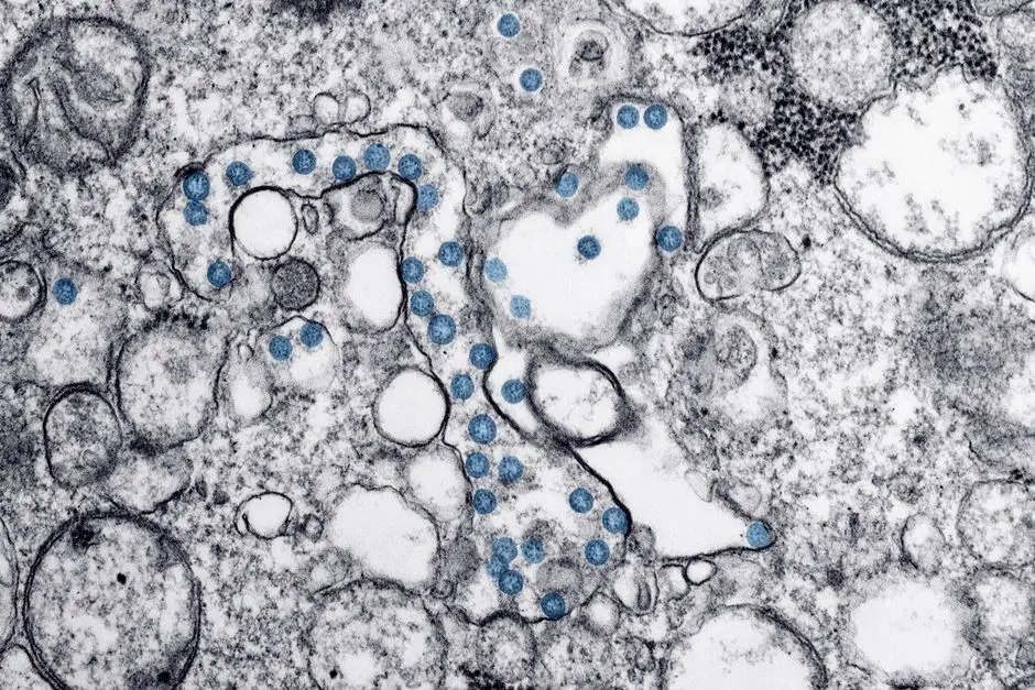
Unlock the secrets of cellular architecture. For decades, the pursuit of finer detail has driven biological discovery, pushing the limits of what our eyes can perceive. While light microscopes opened the initial windows into the microcosm, their inherent limitations, governed by the physics of visible light, prevented us from truly deciphering the intricate machinery within cells. Today, electron microscopes stand as monumental achievements in scientific imaging, shattering previous resolution barriers and revealing structures at an unprecedented nanometer scale.
We embark on an essential journey to dissect the fundamental principles empowering electron microscopes to achieve such superior resolution. We will systematically explore the wave-particle duality of electrons, the critical role of wavelength in defining resolution limits, and the ingenious engineering behind electromagnetic lenses. This deep dive is indispensable for any serious practitioner navigating the complex landscape of biological imaging, providing actionable insights into why these instruments are irreplaceable for groundbreaking research. Master the core concepts that differentiate electron microscopy, and elevate your understanding of advanced laboratory imaging methods for biological structures, crucial for unlocking new biological frontiers.
The Quantum Leap in Detail: Wavelength as the Resolution Determinant
We must first confront the immutable laws governing resolution: the ability to distinguish two closely spaced points as separate entities. This critical metric is fundamentally constrained by the wavelength of the illuminating radiation. Ernst Abbe’s seminal diffraction limit, a cornerstone of optics, dictates that the maximum resolution achievable, d, is approximately half the wavelength (λ) of the light used, divided by the numerical aperture (NA) of the objective lens. The formula, d = λ / (2NA), reveals an inescapable truth: shorter wavelengths yield higher resolution. For visible light, with wavelengths spanning approximately 400 to 700 nanometers (nm), even an ideally constructed light microscope operating with a high NA is inherently restricted to resolving features no smaller than about 200-250 nm. This physical barrier means that intricate subcellular organelles like ribosomes (typically 20-30 nm), individual protein complexes, or the intricate folds of mitochondrial cristae (around 10-20 nm thickness) remain largely indistinct blurs, beyond the reach of conventional light microscopy.
Electron microscopes decisively circumvent this intrinsic limitation by replacing photons with electrons as the imaging medium. While electrons are classically understood as particles, quantum mechanics reveals their wave-like properties. The critical principle underpinning electron microscopy's superior resolution lies in Louis de Broglie’s hypothesis: any moving particle possesses a characteristic wavelength inversely proportional to its momentum. By accelerating electrons to extremely high velocities – often approaching a significant fraction of the speed of light – we drastically reduce their associated de Broglie wavelength. For electrons accelerated by common voltages (e.g., 100-200 kV), the de Broglie wavelength can be as short as 0.003 to 0.008 nm. This represents a reduction by a factor of over 50,000 to 100,000 compared to visible light. This dramatic shortening of the effective wavelength is the primary, foundational reason electron microscopes can resolve structures down to the atomic scale, forging a path to unprecedented detail and revealing the intricate nano-architecture of biological systems.
Harnessing Electron Waves: Acceleration and Focusing Mechanisms
To fully exploit the incredibly short de Broglie wavelengths of electrons, we must implement sophisticated mechanisms for their generation, precise acceleration, and meticulous manipulation. An electron microscope begins with an electron gun, the source of our imaging probe, typically using thermionic or field emission. The generated electrons are then subjected to an immense electric potential difference, often ranging from 20,000 volts (20 kV) to several hundred thousand volts (up to 300 kV) in advanced research microscopes. This high accelerating voltage directly imparts significant kinetic energy to the electrons, propelling them to velocities approaching a substantial fraction of the speed of light. Crucially, this high velocity drives the dramatic reduction in their de Broglie wavelength, directly impacting achievable resolution. We literally forge ultra-short wavelengths by pushing electron speeds to their practical limits, achieving resolutions far beyond the light microscope’s reach.
Once accelerated, these electron waves must be precisely focused and steered, mirroring the function of glass lenses. However, electrons interact strongly with air and optical glass, rendering conventional lenses useless. Instead, electron microscopes employ finely tuned electromagnetic lenses – essentially precisely wound coils of wire generating powerful, symmetrical magnetic fields – to converge and diverge the electron beam. These lenses operate on the fundamental principle that a magnetic field exerts a force on a moving charge, altering its trajectory. By meticulously controlling the current flowing through these coils, we can precisely adjust the strength of the magnetic field, shaping the electron beam. This allows us to guide the electrons through the vacuum column, focus them onto the specimen, and project the magnified image onto a detector. The design and stability of these electromagnetic lens systems are paramount; achieving atomic-scale resolution demands unparalleled geometric accuracy and electronic stability to minimize aberrations and ensure a coherent, tightly focused electron beam, enabling us to dissect the nano-architecture of biological systems with surgical precision.
Overcoming Imperfections: Aberrations and Vacuum Demands
Even with their inherent advantages, electron microscopes face unique challenges, primarily related to lens imperfections and the necessity of a high vacuum environment. Just like optical lenses, electromagnetic lenses are susceptible to aberrations that degrade image quality and limit achievable resolution. The two most significant types are spherical aberration and chromatic aberration. Spherical aberration arises because electrons traveling farther from the optical axis are focused more strongly, leading to a blurred point focus. Chromatic aberration occurs because not all electrons in the beam possess exactly the same energy (and thus velocity), causing electrons of different energies to be focused at slightly different points. These aberrations are far more pronounced in magnetic lenses than in glass lenses, and they historically set a practical limit on resolution even with extremely short electron wavelengths. Modern electron microscopes mitigate these issues through meticulous lens design, very stable high-voltage power supplies, and increasingly, by employing advanced aberration correctors that computationally compensate for lens imperfections, pushing resolution limits further into the sub-ångström regime.
A second, non-negotiable requirement for electron microscopy is an ultra-high vacuum environment within the entire electron column. This isn't merely a convenience; it's a fundamental necessity driven by the nature of electrons. Electrons interact readily with gas molecules, meaning any air molecules present in the column would scatter the electron beam, causing significant signal loss, blurring, and ultimately preventing image formation. We maintain vacuums typically ranging from 10-4 Pascals in the specimen chamber to 10-7 Pascals in the electron gun area. Achieving and maintaining such extreme vacuum levels demands sophisticated pumping systems (rotary, diffusion, turbomolecular, ion pumps) and meticulous sealing of all components. This vacuum requirement also imposes significant constraints on sample preparation, as biological specimens must be dehydrated and often coated to withstand this harsh environment, which can introduce artifacts. Understanding these limitations is crucial for interpreting EM images accurately and for designing experiments that minimize their impact on the biological structures under investigation.
The Art of Seeing Small: Sample Preparation and Contrast Enhancement
Achieving superior resolution in electron microscopy extends far beyond the instrument itself; it critically hinges on meticulous sample preparation, which is an art form in biological electron microscopy. Unlike light microscopy, where live, hydrated samples are often studied, biological specimens for electron microscopy must endure a harsh vacuum and the electron beam's destructive energy. The primary challenge is to preserve cellular ultrastructure faithfully while simultaneously introducing sufficient contrast for electron visualization. Biological matter, composed primarily of light elements (C, H, O, N), is largely transparent to electrons. Therefore, we must impregnate the sample with heavy metals. This process typically involves several key steps: fixation using chemicals like glutaraldehyde and osmium tetroxide to rapidly cross-link proteins and lipids, thereby stabilizing cellular components. Subsequently, dehydration replaces water with organic solvents, a critical step before embedding. Then, embedding in a resin polymerizes to create a solid block, allowing for ultra-thin sectioning.
The next vital step is sectioning: using an ultramicrotome with a diamond or glass knife to cut sections typically 50-100 nanometers thick. This thinness is paramount for electron penetration and minimizing overlapping structures that would obscure detail. Finally, post-staining applies additional heavy metal salts, such as uranyl acetate and lead citrate, to selectively bind to specific cellular components. These heavy metal atoms, with their high atomic number, effectively scatter electrons, creating electron-dense regions that appear dark in the final image, thus providing the necessary contrast. Without these staining agents, most biological structures would be invisible. While these extensive preparation protocols are indispensable for high-resolution imaging, they also represent a significant potential source of artifacts. Recognizing and differentiating between genuine biological features and preparation-induced alterations is a hallmark of an expert electron microscopist. We must consistently evaluate our protocols to ensure the integrity of the biological information gleaned.
Pushing the Boundaries: Advanced Electron Microscopy Techniques
The quest for higher resolution and deeper biological insight continues to drive innovation in electron microscopy, giving rise to specialized techniques that extend its capabilities far beyond conventional imaging. One of the most transformative advancements is Cryo-Electron Microscopy (Cryo-EM). Instead of chemical fixation and dehydration, Cryo-EM involves flash-freezing biological samples in a thin layer of vitreous ice, preserving molecules in their near-native, hydrated state. This revolutionary approach minimizes preparation artifacts, allowing for the determination of protein structures at near-atomic resolution. The ability to image macromolecules without crystallization has opened vast new avenues in structural biology, earning its pioneers a Nobel Prize. We now routinely reconstruct intricate protein complexes, viral particles, and even cellular organelles from thousands of 2D images, leveraging computational power to create stunning 3D models.
Another powerful technique is Electron Tomography (ET), which extends 2D electron micrographs into 3D reconstructions of cellular volumes. By acquiring a series of 2D images of a tilted specimen from multiple angles, sophisticated algorithms can computationally back-project these images to generate a comprehensive 3D model. This allows us to visualize the spatial arrangement of organelles, cytoskeletal networks, and molecular machines within intact cells or thicker sections. Furthermore, advancements in Scanning Transmission Electron Microscopy (STEM) and aberration-corrected lenses allow for routine atomic-resolution imaging, even for biological samples, providing unprecedented insight into material properties and single-atom localization within biological contexts. These advanced techniques exemplify our relentless pursuit of detail, continually pushing the frontiers of resolution and enabling us to unravel the most complex biological puzzles with precision and clarity previously unimaginable.

Navigating the Nanoworld: Best Practices and Common Pitfalls
To consistently extract high-resolution information from electron microscopes, we must adhere to rigorous best practices and remain vigilant against common pitfalls. Achieving optimal resolution is a holistic endeavor, demanding excellence at every stage, from sample preparation to image acquisition and interpretation. For sample preparation, consistency is paramount. Deviations in fixation times, buffer molarities, or dehydrating agent concentrations can introduce subtle artifacts that compromise structural integrity. Always process controls alongside experimental samples to confidently distinguish biological variation from preparation artifacts. Furthermore, maintaining an immaculate laboratory environment for sectioning and grid handling is crucial; dust particles, fingerprints, or residual chemicals on grids can severely degrade image quality, often masking the very details we strive to resolve.
During image acquisition, optimal instrument alignment is non-negotiable. Regular calibration of lens currents, astigmatism correction, and precise focus adjustment directly impact the fidelity of the final image. We must consistently monitor and compensate for specimen drift, which, even at nanometer scales, can severely blur high-resolution details. A common pitfall for new users is over-exposing the sample to the electron beam, leading to radiation damage and structural degradation. Learn to balance signal-to-noise ratio with minimal dose exposure, especially for sensitive biological specimens. Finally, in image interpretation, resist the urge to over-interpret faint signals or assume perfect representation. Correlate EM findings with data from other imaging modalities whenever possible to build a robust understanding. Forge a critical eye, question assumptions, and continually refine your methodological approach to unlock the full potential of electron microscopy and ensure the integrity of your biological discoveries.
Key Takeaways
De Broglie Wavelength: The Resolution Multiplier
Electron microscopes achieve superior resolution by harnessing the wave-like properties of electrons. Accelerating electrons to high velocities dramatically shortens their de Broglie wavelength, enabling the visualization of structures far smaller than the 200 nm limit of visible light. This fundamental principle—shorter wavelength, higher resolution—is the bedrock of EM’s power.
Electromagnetic Lenses: Precision Beam Control
Unlike light, electrons are focused by powerful electromagnetic lenses, not glass. These precisely controlled magnetic fields guide, converge, and steer the electron beam, crucial for forming high-magnification, high-resolution images. The accuracy and stability of these lenses are paramount for minimizing aberrations.
Vacuum & Aberrations: Technical Demands and Solutions
A high vacuum environment is essential to prevent electron scattering by gas molecules. Additionally, electromagnetic lenses are prone to aberrations (spherical, chromatic) that can blur images. Modern EM designs and aberration correctors actively mitigate these imperfections, pushing resolution to sub-ångström levels.
Meticulous Sample Preparation: Unlocking Contrast
Biological samples require extensive preparation—fixation, dehydration, embedding, ultra-thin sectioning, and heavy metal staining—to withstand the vacuum and electron beam, and to introduce the necessary electron contrast. This intricate process is vital for revealing ultrastructural details.
Advanced EM: Cryo-EM and 3D Insights
Techniques like Cryo-EM preserve samples in their near-native state, revolutionizing structural biology by allowing atomic-resolution imaging of macromolecules without crystallization. Electron Tomography provides 3D reconstructions of cellular volumes, offering spatial context to ultrastructural details.
FAQ
-
What is the primary reason electron microscopes offer higher resolution than light microscopes?
The primary reason is the significantly shorter wavelength of electrons compared to visible light. According to Abbe's diffraction limit, resolution is directly proportional to wavelength. By accelerating electrons, their de Broglie wavelength can be thousands of times shorter than light, enabling the visualization of structures at the nanometer scale.
-
How do electron microscopes focus electrons if they cannot use glass lenses?
Electron microscopes utilize electromagnetic lenses, which are coils of wire that generate strong magnetic fields. These magnetic fields exert a force on the moving electrons, causing them to converge or diverge, thereby focusing the electron beam in a manner analogous to how glass lenses focus light.
-
Why is sample preparation for electron microscopy so complex for biological specimens?
Biological samples are typically soft, hydrated, and mostly transparent to electrons. They require complex preparation steps—including chemical fixation, dehydration, embedding in resin, ultra-thin sectioning, and heavy metal staining—to preserve their delicate ultrastructure, withstand the high vacuum, and introduce sufficient electron contrast for imaging.