
Uncover Cellular Secrets: Advanced Laboratory Imaging Methods for Biological Structures

In the relentless pursuit of understanding life's intricate machinery, advanced laboratory imaging methods stand as indispensable pillars. These sophisticated techniques empower biologists to peer into the microscopic world, revealing the delicate architectures of cells, the dynamic interplay of biomolecules, and the complex choreography of biological processes. From unraveling the static beauty of cellular ultrastructure to capturing the living ballet within a cell, mastering these imaging methodologies is paramount for any serious biological inquiry.
This comprehensive guide dives deep into the arsenal of cutting-edge imaging tools, dissecting their principles, applications, and practical nuances. We forge a pathway through the optical and electron realms, illuminating how each technique contributes uniquely to our cumulative biological knowledge. Prepare to elevate your understanding and wield these powerful instruments with precision, transforming unseen biological realities into profound scientific insights.
Forging the Vision: Foundations of Light Microscopy in Biological Research
We initiate our exploration by grounding ourselves in the foundational principles that govern the visualization of life: light microscopy. This venerable technique remains the cornerstone of biological imaging, offering unparalleled accessibility and versatility for scrutinizing cellular and tissue architecture. Understanding the fundamental principles of how light microscopy operates in biological research is crucial; it involves the interaction of light with biological samples, generating magnified images through a series of lenses. We manipulate wavelength, aperture, and illumination to optimize contrast and resolution, transforming transparent biological entities into observable phenomena.
The diversity within this field is vast. We recognize the main categories of light microscopy available including brightfield, phase contrast, differential interference contrast (DIC), darkfield, and polarization microscopy. Each variant capitalizes on different optical properties of light, allowing us to highlight specific features or overcome limitations inherent in transparent biological specimens. For instance, while brightfield microscopy is straightforward, relying on differential absorption of light, it often requires staining. Conversely, we navigate the subtleties required for unstained samples; here, understanding a clear explanation of brightfield versus phase contrast microscopy becomes indispensable. Phase contrast, developed by Frits Zernike, converts phase shifts in light passing through a sample into amplitude differences, thereby enhancing contrast in transparent, living cells without the need for toxic stains. This allows us to observe cellular processes in their natural state, a distinct advantage over methods requiring fixation. We meticulously select the correct technique to match our experimental objectives, ensuring optimal data acquisition.
Optimizing Observation: Applications and Selection in Optical Microscopy
Leveraging light microscopy effectively requires a strategic understanding of its diverse applications and the judicious selection of instrumentation. We deploy optical microscopy across a spectrum of biological investigations, from routine cell culture monitoring to intricate histological analyses. The the diverse applications of optical microscopy within biology laboratories span cell counting, morphology assessment, parasite identification, tissue pathology, and even basic developmental studies. It provides an immediate, cost-effective window into biological samples, often serving as the first line of investigation before more complex techniques are employed.
However, successful imaging isn't merely about having a microscope; it's about making an informed choice. We meticulously consider essential considerations when selecting the appropriate microscope for specific biological samples. This decision hinges on several critical factors: the nature of the sample (live vs. fixed, stained vs. unstained), the desired magnification and resolution, the specific biological question (morphology, dynamics, localization), and budget constraints. For instance, examining bacterial motility demands a phase contrast or darkfield microscope, while observing stained tissue sections might favor brightfield. We also assess objective lenses (magnification, numerical aperture, working distance), illumination sources (halogen, LED), and camera systems (monochrome for sensitivity, color for histological staining). Errors in selection can lead to suboptimal data, missed observations, or unnecessary expenditure. We champion a 'fit-for-purpose' approach, ensuring that our chosen tool precisely aligns with our investigative needs, thereby maximizing the scientific return on investment and propelling our discoveries forward.
Igniting Insights: Principles of Fluorescence Microscopy for Biomolecule Visualization
We elevate our imaging capabilities by transitioning to fluorescence microscopy, a technique that has revolutionized our ability to visualize specific biomolecules within the complex cellular milieu. Unlike conventional light microscopy that relies on light absorption or scattering, fluorescence microscopy exploits the phenomenon where specific molecules, called fluorophores, absorb light at one wavelength and emit it at a longer, lower-energy wavelength. We harness the intricate mechanism by which fluorescence microscopy visualizes biomolecules by attaching these fluorophores to target structures, such as proteins, DNA, or organelles. An excitation light source (often a mercury lamp or LED) illuminates the sample, causing the fluorophores to emit light. Filters then separate this emitted light from the excitation light, allowing only the fluorescent signal to reach the detector.
The utility of fluorescence microscopy extends across virtually every domain of modern biology. We consistently ask, the primary applications of fluorescence microscopy? Its power lies in its specificity and sensitivity, enabling us to pinpoint the localization of proteins, quantify gene expression, track cellular processes, visualize subcellular compartments, and even detect specific pathogens. The advent of genetically encoded fluorescent proteins like GFP (Green Fluorescent Protein) has been particularly transformative, allowing us to tag and visualize molecules in living cells without external staining. Crucially, the quality and type of fluorescent labels are paramount. We depend heavily on the pivotal role of fluorescent dyes and markers in effective cell imaging. These include synthetic organic dyes (e.g., DAPI for DNA, Phalloidin for actin), immunofluorescence (antibodies conjugated to fluorophores), and the aforementioned genetically encoded reporters. Selecting the right fluorophore involves considering its excitation/emission spectra, photostability, brightness, and compatibility with the experimental system, ensuring we capture the clearest, most relevant biological signals.
Precision Preparation: Sample Handling and Troubleshooting in Fluorescence Imaging
The success of fluorescence microscopy hinges significantly on meticulous sample preparation and a proactive approach to troubleshooting. We understand that a brilliantly designed experiment can yield misleading results if the sample is not correctly handled. Therefore, mastering the critical steps involved in preparing samples for fluorescence microscopy is non-negotiable. For fixed cells, this involves careful fixation (e.g., paraformaldehyde to preserve morphology), permeabilization (e.g., Triton X-100 to allow antibody penetration), blocking (to prevent non-specific binding), and incubation with primary and secondary antibodies (if using immunofluorescence) or direct staining with fluorescent dyes. For live-cell imaging, we prioritize maintaining physiological conditions, minimizing phototoxicity, and ensuring proper mounting to prevent drift. Key considerations include choosing appropriate media, temperature control, and CO2 regulation. Any compromise in these stages can lead to artifacts, diminished signal, or cell damage, severely impacting data integrity.
Even with impeccable preparation, challenges invariably arise. We must vigilantly identify and mitigate how to identify and mitigate common imaging artifacts encountered in fluorescence microscopy. These can range from background fluorescence due to non-specific antibody binding or autofluorescence from the sample itself (e.g., NADH, flavins), to photobleaching (loss of fluorescence intensity over time due to light exposure) and chromatic aberration (different wavelengths focusing at different points). We employ strategies such as optimizing antibody concentrations, using autofluorescence-reducing reagents, adding anti-fade mounting media, and employing sequential excitation for multi-channel imaging to combat photobleaching and crosstalk. Furthermore, understanding the impact of optical components, such as dirty objectives or misaligned filters, is crucial. Proactive troubleshooting, combining a systematic diagnostic approach with a deep understanding of microscopy principles, empowers us to acquire robust, reliable, and biologically meaningful fluorescent data.
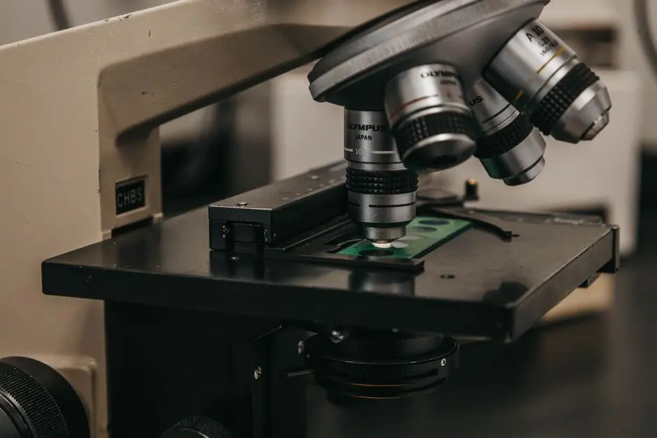
Sharpening the View: How Confocal Microscopy Refines Cellular Imaging
While conventional fluorescence microscopy offers unparalleled specificity, it suffers from out-of-focus blur, particularly in thicker samples. We transcend this limitation with confocal microscopy, a revolutionary technique designed to improve image quality and enable optical sectioning. the transformative ways confocal microscopy enhances cellular imaging capabilities lies in its ability to collect light from a single focal plane, effectively rejecting out-of-focus fluorescence. This provides dramatically clearer images of cellular structures, even deep within tissues. We achieve this precision through a pinhole aperture placed in the detection pathway, optically conjugating with the focal plane of the objective lens. This strategic aperture blocks emitted light from above and below the focal plane, ensuring only in-focus photons contribute to the final image.
Understanding the defining characteristics of confocal laser scanning microscopy reveals its operational elegance. A laser scans the sample point by point, line by line, building up an image pixel by pixel. This precise control over illumination and detection confers significant the significant advantages confocal microscopy offers over traditional imaging methods. We achieve superior spatial resolution, particularly in the Z-axis, and significantly improved signal-to-noise ratio. This empowers us to generate high-contrast, high-resolution images of thick biological samples, minimizing the impact of scattered light. Consequently, the impactful applications of confocal microscopy in contemporary biology research are vast, including 3D reconstruction of cells and tissues, colocalization studies of multiple proteins, analysis of dynamic cellular processes (e.g., calcium signaling), and imaging of live organisms. The cornerstone of its power is the sophisticated process of optical sectioning in confocal imaging. By acquiring a series of images at different focal depths (Z-stacks), we reconstruct a three-dimensional representation of the sample, providing an unprecedented perspective on cellular architecture and organization. This ability to digitally dissect a specimen without physical slicing is a game-changer for spatial biology.
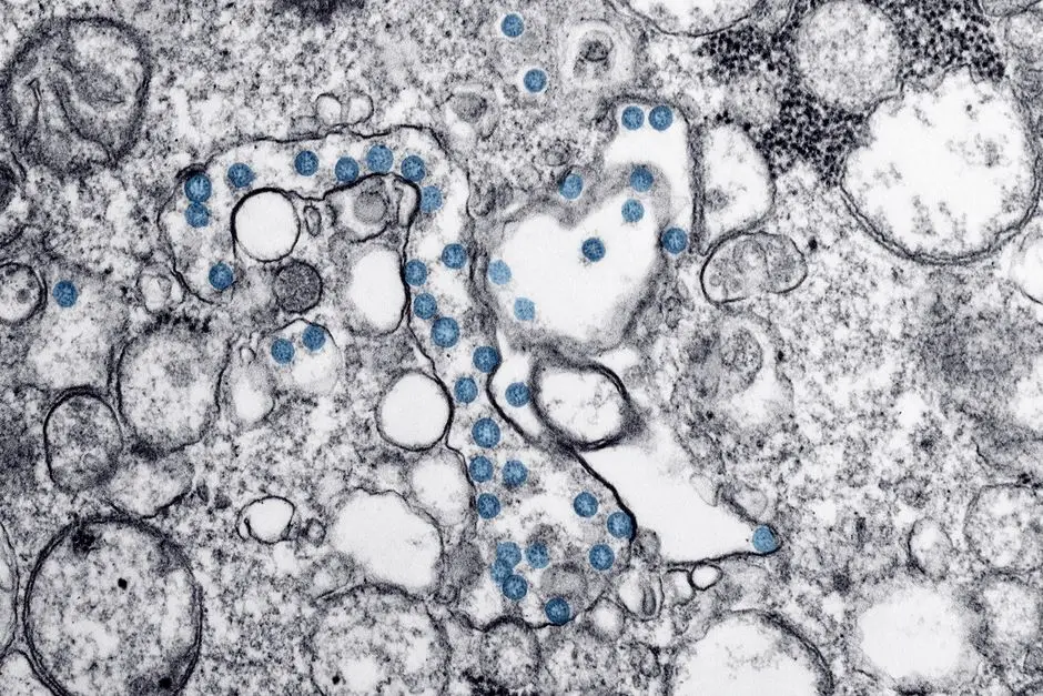
Unveiling Ultrastructure: The Power of Electron Microscopy
When our quest demands resolution far beyond the limits of light, we turn to electron microscopy. This powerful suite of techniques allows us to visualize the intricate ultrastructure of cells and tissues at nanometer scale, revealing details that are simply invisible with light. how electron microscopy uniquely reveals cellular ultrastructure with unparalleled detail centers on its use of a beam of electrons instead of photons. Due to the much shorter wavelength of electrons compared to light, electron microscopes achieve significantly higher resolution, enabling us to observe organelles, macromolecular complexes, and even individual proteins within a cell. The electrons interact with the specimen, producing signals that are then captured and converted into an image.
A fundamental distinction exists between the primary modalities. We delineate the critical distinctions between Transmission Electron Microscopy (TEM) and Scanning Electron Microscopy (SEM). TEM passes electrons through ultrathin sections of a sample, revealing internal structures and subcellular organization, akin to an X-ray of the cell. SEM, conversely, scans the surface of a sample with a focused electron beam, generating a highly detailed 3D topographic image of the specimen's exterior. Both require specialized preparation. We rigorously master the specialized protocols for preparing biological samples for electron microscopy, which involve fixation (e.g., glutaraldehyde, osmium tetroxide), dehydration, embedding in resin, and sectioning (for TEM) or critical point drying and metal coating (for SEM). These steps are crucial for preserving cellular integrity and enhancing contrast. The the profound applications of electron microscopy in molecular biology studies are diverse, from identifying viral particles and characterizing cellular junctions to elucidating protein complex structures and diagnosing diseases at the ultrastructural level. The fundamental reason the fundamental reasons why electron microscopes achieve significantly higher resolution lies in the wave-particle duality of electrons. The de Broglie wavelength of an accelerated electron is orders of magnitude smaller than that of visible light, bypassing the diffraction limit that constrains optical microscopes. This enables us to resolve structures down to fractions of a nanometer, pushing the boundaries of biological discovery.
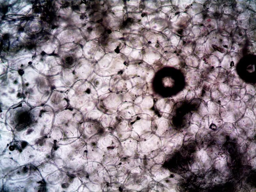
Capturing Dynamics: Live-Cell Imaging and Time-Lapse Microscopy
While fixed-cell imaging provides snapshots of biological reality, understanding dynamic processes requires us to observe living cells in real-time. This is the domain of live-cell imaging, a methodology that empowers us to witness biology in motion. We strategically employ the innovative methods by which live-cell imaging tracks dynamic biological processes by maintaining cells under physiological conditions while continuously acquiring images over extended periods. This allows us to observe phenomena such as cell division, migration, intracellular transport, protein trafficking, and responses to stimuli as they unfold, providing invaluable kinetic data.
Successful live-cell imaging demands specialized instrumentation and careful experimental design. We must ensure we have the essential equipment required for successful live-cell microscopy setups. This typically includes an inverted microscope (for imaging cells in dishes), an environmental chamber (to maintain temperature, CO2, and humidity), a highly sensitive camera, and stable motorized stages for long-term observation. We meticulously select objectives with low phototoxicity and high light transmission. Beyond hardware, we implement various various effective techniques for imaging living cells over extended periods. These include time-lapse imaging (sequential image acquisition), FRAP (Fluorescence Recovery After Photobleaching) to study protein mobility, FRET (Förster Resonance Energy Transfer) for molecular interactions, and calcium imaging for signaling events. Each technique offers a unique window into specific cellular dynamics. However, we acknowledge the the inherent challenges often encountered in live-cell imaging experiments. These include phototoxicity (damage to cells from prolonged light exposure), photobleaching (loss of fluorescence signal), maintaining cell viability and focus over long durations, and managing large datasets. Mitigation strategies involve using low-light illumination, high-sensitivity detectors, and specialized cell culture conditions. Despite these hurdles, the the diverse applications of time-lapse microscopy across various biological disciplines are transformative, providing insights into developmental biology, neurobiology, immunology, cancer research, and drug discovery. By capturing the kinetic essence of life, we uncover the mechanisms that drive cellular behavior and disease progression.
Integrating and Innovating: Future Perspectives in Biological Imaging
As we survey the vast landscape of laboratory imaging methods, it becomes clear that no single technique offers a complete picture. The true power often resides in the intelligent integration of these tools and the relentless pursuit of innovation. We actively seek to combine modalities, leveraging the strengths of each. For instance, correlative light and electron microscopy (CLEM) allows us to pinpoint fluorescently labeled structures with light microscopy and then examine their ultrastructure with electron microscopy, bridging the resolution gap between the two domains. This synergistic approach maximizes the information harvested from precious biological samples.
The field is in a constant state of evolution. We witness the emergence of super-resolution microscopy techniques (e.g., STED, PALM, STORM), which shatter the classical diffraction limit of light, pushing optical resolution down to tens of nanometers, rivaling some electron microscopy capabilities for specific applications. These innovations allow us to visualize molecular interactions and arrangements within cells with unprecedented clarity, bringing us closer to understanding molecular machines in their native environment. Furthermore, advancements in computational imaging, AI-driven image analysis, and automated microscopy platforms are transforming how we acquire, process, and interpret vast quantities of image data. Machine learning algorithms now assist in object segmentation, tracking, and even phenotype classification, accelerating discovery and reducing subjective biases. We champion an adaptive mindset, continuously exploring novel probes, smarter optics, and more powerful computational tools. Our commitment to these integrated and innovative strategies will continue to unlock deeper biological mysteries, propelling us towards a comprehensive understanding of life's intricate structures and dynamic processes at every scale.
Key Takeaways
Light Microscopy: The Foundation
Light microscopy remains critical for initial observations, offering diverse techniques like brightfield, phase contrast, and DIC. It visualizes samples based on light interaction and is essential for basic cell morphology and tissue analysis. Choosing the right light microscope involves considering sample type, desired resolution, and experimental goals.
Fluorescence Microscopy: Specificity and Sensitivity
Fluorescence microscopy leverages fluorophores to specifically label and visualize biomolecules. It's vital for protein localization, gene expression, and tracking cellular processes. Effective use requires meticulous sample preparation to avoid artifacts like photobleaching and autofluorescence, ensuring clear, biologically relevant signals.
Confocal Microscopy: Enhanced Spatial Resolution and 3D Imaging
Confocal microscopy improves upon traditional fluorescence by using a pinhole to reject out-of-focus light, providing superior spatial resolution, especially in the Z-axis. This enables optical sectioning and 3D reconstruction of cells and tissues, crucial for complex structural and dynamic studies within thicker samples.
Electron Microscopy: Unveiling Ultrastructure
Electron microscopy (TEM and SEM) utilizes electron beams for ultra-high resolution imaging, revealing cellular ultrastructure at nanometer scales. TEM visualizes internal structures, while SEM offers detailed surface topography. Specialized sample preparation is mandatory to preserve integrity and enhance contrast for these techniques, crucial for molecular biology and detailed pathology.
Live-Cell Imaging: Dynamics in Real-Time
Live-cell imaging tracks biological processes in real-time under physiological conditions, providing kinetic data on cell division, migration, and molecular dynamics. It requires specialized equipment (environmental chambers, sensitive cameras) and addresses challenges like phototoxicity and focus drift. This technique is indispensable for understanding dynamic cellular behaviors and responses.
Integration and Innovation: The Future of Biological Imaging
The field evolves through integrating diverse techniques (e.g., correlative light and electron microscopy) and embracing innovations like super-resolution microscopy and AI-driven image analysis. Combining approaches maximizes information, pushes resolution limits, and enhances data interpretation, continuously advancing our understanding of biological systems.
FAQ
-
What is the primary advantage of fluorescence microscopy over brightfield microscopy?
The primary advantage of fluorescence microscopy is its specificity and sensitivity. It allows us to selectively label and visualize specific biomolecules (e.g., proteins, DNA) within a complex cellular environment, providing high contrast against a dark background. Brightfield microscopy, in contrast, visualizes structures based on light absorption and typically requires staining, which can lack specificity and often kills live cells.
-
When should I choose electron microscopy instead of light microscopy?
You should choose electron microscopy when your research question demands significantly higher resolution to visualize cellular ultrastructure, organelles, or macromolecular complexes that are below the diffraction limit of light microscopy (typically <200 nm). Electron microscopy can achieve resolutions down to fractions of a nanometer, making it essential for detailed structural analysis at the subcellular level.
-
What are the main challenges in live-cell imaging, and how can they be mitigated?
Main challenges in live-cell imaging include phototoxicity (damage to cells from light exposure), photobleaching (loss of fluorescence signal), maintaining physiological conditions (temperature, CO2, humidity) for cell viability, and focus drift. Mitigation strategies include using low-intensity light sources, highly sensitive cameras, anti-fade reagents, environmental chambers, automated focus systems, and selecting robust cell lines or specific fluorescent probes that are less prone to phototoxicity and photobleaching.