
> Laboratory Techniques > Microscopy and Cellular Imaging > Optimize Fluorescence Imaging: Detect and Resolve Common Artifacts
Optimize Fluorescence Imaging: Detect and Resolve Common Artifacts
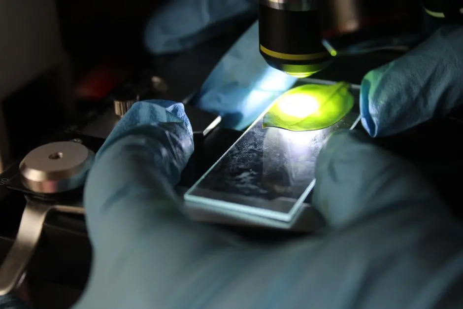
Fluorescence microscopy unveils the hidden architectures and dynamic processes within biological systems with unparalleled resolution. Yet, its transformative power is often compromised by elusive imaging artifacts—phantom signals that distort data, mislead interpretations, and undermine quantitative analyses. This deep dive equips researchers, from novices to seasoned investigators, with the surgical precision required to identify, understand, and surgically eliminate these common pitfalls. Our collective mission is to elevate image integrity, allowing us to accurately decipher cellular dynamics and structural nuances. We forge a path towards pristine visualization, critically examining how to overcome challenges inherent in the advanced methodologies of biological imaging that underpin modern discovery. Prepare to transform your approach, translating raw data into robust, publication-ready imagery and solidifying the foundation of your biological insights.
The Imperative of Artifact Management in Fluorescence Microscopy
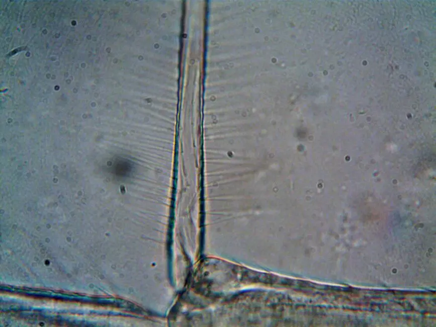
We embark on a critical exploration: understanding why imaging artifacts represent more than mere visual nuisances—they are fundamental threats to scientific veracity. In fluorescence microscopy, an artifact is any feature in an image that does not originate directly from the sample's true biological signal but arises from the instrumentation, sample preparation, or environmental conditions. Ignoring these distortions leads to misinterpretation of cellular events, erroneous quantification, and ultimately, flawed conclusions. We must cultivate a keen eye for these spectral shadows and optical illusions.
Our analytical journey begins with recognizing that every component of the imaging system, from the light source and fluorophores to the optical path and detector, can introduce discrepancies. For instance, an improperly calibrated system might generate uneven illumination, falsely suggesting variations in fluorophore concentration. Or, a poorly mounted sample could induce optical aberrations, blurring critical subcellular details. We must adopt a proactive, systematic approach to imaging, treating each acquisition as a diagnostic challenge. By rigorously scrutinizing every image for tell-tale signs of artifacts, we transition from passive observers to active interrogators of our data. This vigilance ensures that the compelling narratives we extract from our images are grounded in biological reality, not instrumental illusion. We optimize not just for visual appeal, but for absolute quantitative integrity, transforming our raw data into irrefutable evidence.
Unmasking Photobleaching and Autofluorescence: Intrinsic Biological Hurdles
We confront two of the most ubiquitous and often intertwined challenges in fluorescence microscopy: photobleaching and autofluorescence. Photobleaching is the irreversible photochemical destruction of a fluorophore due to excessive exposure to excitation light. This phenomenon diminishes signal intensity over time, particularly critical in live-cell imaging or extended time-lapse experiments. We observe a reduction in fluorescence, which can be mistakenly interpreted as a decrease in biological concentration or activity. To combat this, we optimize by reducing excitation power, shortening exposure times, utilizing highly photostable fluorophores, and incorporating anti-fade reagents into our mounting media. Crucially, we consider environmental factors, such as oxygen concentration, which can accelerate photobleaching.
Autofluorescence, on the other hand, originates from the natural emission of light by intrinsic cellular components—molecules like NADH, FAD, lipofuscins, collagen, and elastin—without the addition of exogenous fluorescent probes. This intrinsic background signal can spectrally overlap with our target fluorophores, obscuring genuine signals and reducing contrast. Its prevalence makes accurate quantification incredibly challenging, especially in tissue samples. We mitigate autofluorescence by carefully selecting fluorophores with emission spectra distinct from common autofluorescent compounds, employing spectral unmixing techniques, and, in some cases, photobleaching the autofluorescence (though this must be done cautiously to avoid damaging the sample). Effective strategies also include specific blocking agents during sample preparation. We navigate these intrinsic hurdles with strategic fluorophore selection and sophisticated imaging techniques, ensuring our insights are sharply focused on the exogenous probes.

Decoding Optical Aberrations: The Lens's Imperfections and Their Impact
We dissect the inherent imperfections within our optical systems—optical aberrations—that profoundly degrade image quality, resolution, and signal fidelity. Among the most critical is spherical aberration, where light rays passing through different regions of the objective lens (paraxial vs. marginal) fail to converge at a single focal point. This results in a blurred image, reduced signal intensity, and a loss of axial resolution. Its primary culprits are refractive index mismatches between the immersion medium, coverslip, and sample, or incorrect coverslip thickness. We surgically address spherical aberration by meticulously matching the refractive index of the immersion oil, using precisely calibrated coverslips, and leveraging objectives equipped with correction collars for fine-tuning. Adaptive optics represent an advanced frontier for dynamic, real-time correction.
Another significant challenge is chromatic aberration, where different wavelengths of light are focused at slightly different points due to the dispersion properties of the lens material. This leads to a colored fringe around objects, especially noticeable at the edges of the field of view, causing misregistration of multi-color images and compromised spatial accuracy. We conquer chromatic aberration by utilizing high-quality apochromatic or super-apochromatic objectives, which are specifically designed to minimize this effect across multiple wavelengths. Post-acquisition image registration can partially compensate for axial and lateral chromatic shifts, but proactive objective selection remains our most potent weapon. By understanding and actively correcting these optical imperfections, we unleash the full theoretical resolution power of our microscopes, pushing the boundaries of what we can resolve within the cellular landscape.
Confronting Electronic Noise and Detector Saturation: The Digital Image Frontier
We navigate the digital realm of image acquisition, where electronic limitations introduce their own set of critical artifacts: electronic noise and detector saturation. Electronic noise manifests as random fluctuations in the detected signal, obscuring genuine biological information, especially at low light levels. We categorize it into several forms: readout noise (inherent to the detector's electronics), shot noise (arising from the quantum nature of light itself, signal-dependent), and dark current noise (thermal generation of electrons in the absence of light). To combat noise, we employ cooled detectors (e.g., scientific CMOS or EMCCD cameras) to minimize dark current, optimize gain settings for maximum signal-to-noise ratio, and utilize signal averaging or binning techniques. We accept shot noise as an irreducible physical limit, focusing instead on maximizing signal detection.
Conversely, detector saturation occurs when the number of photons hitting a pixel exceeds its maximum storage capacity, causing the pixel value to 'clip' at its highest digital value (e.g., 255 for an 8-bit image, 4095 for a 12-bit image). Saturated pixels lose all quantitative information, appearing as uniformly bright 'blobs' without internal detail. This artifact frequently arises from excessively high excitation power or prolonged exposure times. We prevent saturation by judiciously adjusting laser intensity, shortening exposure duration, or incorporating neutral density (ND) filters into the excitation path. Crucially, we monitor the pixel intensity histogram during acquisition, ensuring that no significant portion of our signal is lost to clipping. Mastering the detector's dynamic range—the ratio between the largest and smallest measurable signal—is paramount for capturing the full spectrum of cellular intensities without compromise, ensuring every photon counts accurately.
Strategic Mitigation: Proactive Measures and Advanced Corrections for Pristine Imaging
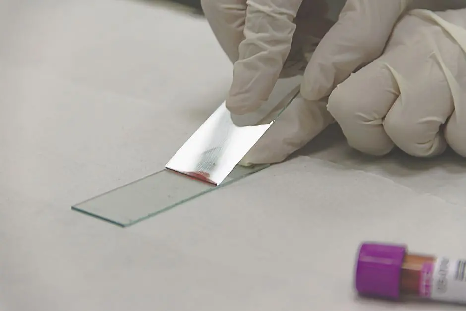
We culminate our exploration by forging a comprehensive strategy for artifact prevention and correction, empowering us to achieve unparalleled image integrity. The battle against artifacts begins long before we press the 'acquire' button. Meticulous sample preparation is paramount: ensuring proper fixation, optimized permeabilization, and precise antibody staining protocols minimize non-specific binding and morphological distortion. The choice of mounting media is critical; it must match the refractive index of the immersion oil and coverslip, and ideally contain anti-fade reagents to curb photobleaching. The quality and thickness of the coverslip are non-negotiable—using precisely specified #1.5H (170 µm) coverslips is essential for minimizing spherical aberration with high NA objectives.
During acquisition, we exercise surgical control over every parameter. Objective selection is not merely about magnification but about numerical aperture (NA), correction for chromatic and spherical aberrations, and compatibility with immersion media. We precisely adjust excitation power and exposure time to maximize signal-to-noise ratio without inducing photobleaching or detector saturation. For multi-color imaging, careful selection of filter sets and sequential acquisition (when appropriate) prevents spectral bleed-through. Post-acquisition, advanced computational methods offer powerful corrective measures. Deconvolution algorithms mathematically reassign out-of-focus light to its point of origin, dramatically enhancing resolution and contrast, particularly in 3D datasets. While selective image processing techniques like background subtraction and denoising can improve visual clarity, we apply them cautiously, especially for quantitative analysis, to avoid introducing new distortions or altering genuine biological signals. Our holistic approach, integrating rigorous preparation with intelligent acquisition and judicious post-processing, unlocks the true potential of fluorescence microscopy.
Key Takeaways
Photobleaching & Autofluorescence: Intrinsic Hurdles
Photobleaching is the irreversible destruction of fluorophores by excitation light, diminishing signal. Combat it with lower laser power, shorter exposures, photostable fluorophores, and anti-fade reagents. Autofluorescence is natural emission from cellular components, obscuring true signals. Mitigate it via distinct fluorophore spectra, spectral unmixing, and specific blocking agents.
Optical Aberrations: Lens Imperfections
Spherical aberration results from refractive index mismatches, blurring images. Correct with proper immersion oil, precise coverslip thickness, and objective correction collars. Chromatic aberration causes different wavelengths to focus at different points, leading to colored fringes. Counteract it with high-quality apochromatic objectives and judicious post-acquisition alignment.
Electronic Noise & Detector Saturation: Digital Limits
Electronic noise (readout, shot, dark current) degrades signal-to-noise ratio. Minimize with cooled detectors, optimized gain, and signal averaging. Detector saturation occurs when pixels are overexposed, losing quantitative data. Prevent it by adjusting excitation power, exposure time, or using neutral density filters, ensuring the full dynamic range is captured.
Strategic Mitigation: Proactive & Corrective Measures
Achieve pristine imaging through a holistic approach. Proactive measures include meticulous sample preparation (fixation, permeabilization), appropriate mounting media, precise coverslip quality, and optimal objective selection. During acquisition, fine-tune laser power, exposure, and filter sets. Post-acquisition, leverage deconvolution for resolution enhancement and cautiously apply image processing for refinement, always prioritizing prevention over correction.
FAQ
-
How do I distinguish an artifact from a real biological signal in my images?
Distinguishing artifacts requires a keen eye and systematic troubleshooting. We look for patterns inconsistent with known biology: uniform speckles across the image (noise), blurred fringes around structures (chromatic aberration), or areas of overexposure lacking detail (saturation). We perform control experiments (e.g., imaging unstained cells for autofluorescence, acquiring images with varying laser power) and cross-reference our observations with published literature and known cellular structures. Reproducibility across multiple samples and imaging sessions also helps confirm a true biological signal versus a sporadic artifact.
-
What is the most common artifact in live-cell imaging and how can I minimize it?
In live-cell imaging, photobleaching stands out as the most common and detrimental artifact, alongside phototoxicity. To minimize photobleaching, we adopt a multi-pronged strategy: reducing excitation light intensity, using highly photostable fluorophores or genetically encoded fluorescent proteins, minimizing exposure times, and employing anti-fade reagents (though less common in live cells due to toxicity). Furthermore, maintaining optimal physiological conditions (temperature, CO2) and using oxygen scavengers (if compatible with the assay) can significantly extend fluorophore lifespan and cell viability during prolonged imaging sessions.
-
Can image processing completely remove artifacts, or should I focus on prevention?
While image processing techniques like deconvolution, background subtraction, and denoising can significantly mitigate the visual impact of certain artifacts, they rarely 'remove' them completely without some risk of data alteration. We prioritize prevention as the cornerstone of robust microscopy. An image acquired with minimal artifacts is inherently more reliable for quantitative analysis than one heavily 'corrected' post-acquisition. Image processing serves as a powerful refinement tool, enhancing data acquired under optimal conditions, but it cannot fully compensate for fundamental issues introduced during sample preparation or acquisition. We strive for pristine data at the source.
-
What role does objective selection play in preventing imaging artifacts?
Objective selection is absolutely critical for artifact prevention. A high-quality objective with appropriate specifications can proactively address several issues. We prioritize objectives with high Numerical Aperture (NA) for maximal light collection and resolution. Crucially, we select objectives corrected for chromatic aberration (apochromatic objectives) to ensure precise color alignment for multi-channel imaging. For samples requiring immersion, we choose objectives compatible with the correct immersion medium (oil, water, glycerol) to avoid spherical aberration, often ensuring the refractive index matches that of our sample and coverslip. Objectives with correction collars also allow us to fine-tune for variations in coverslip thickness or sample depth. This strategic choice of objective forms a foundational defense against optical artifacts.