
> Laboratory Techniques > Microscopy and Cellular Imaging > Master Confocal Microscopy: Advanced Cellular Imaging
Master Confocal Microscopy: Advanced Cellular Imaging
In the relentless pursuit of biological understanding, our capacity to visualize the intricate architecture and dynamic processes within living systems dictates the pace of discovery. Traditional widefield microscopy, while foundational, often falls short, yielding images blurred by out-of-focus light and obscuring the true depth of cellular structures. This inherent limitation has long challenged researchers striving for clarity at the subcellular level.
We stand at a critical juncture where pushing the boundaries of optical imaging is not merely advantageous but imperative. This comprehensive resource dissects the profound impact of Confocal Laser Scanning Microscopy (CLSM), a revolutionary technique that has transformed our ability to probe biological phenomena with unprecedented precision. CLSM stands out among the most sophisticated Laboratory Imaging Methods for Biological Structures available today, empowering scientists to generate stunning, high-resolution 3D reconstructions of cells and tissues. We shall forge a deep understanding of its core principles, dissect its crucial components, unveil its myriad applications, and navigate the common pitfalls to optimize your experimental outcomes. Prepare to unlock the strategic insights that will elevate your imaging prowess and propel your biological investigations forward.
Deconstruct Confocal Microscopy: The Core Principle
Confocal Laser Scanning Microscopy (CLSM) represents a paradigm shift from conventional widefield fluorescence microscopy, fundamentally by eliminating out-of-focus light. This distinction is paramount. In widefield microscopy, the entire sample is illuminated, and all emitted fluorescence from the specimen, regardless of its focal plane, contributes to the final image. The result is often a blurry overlay of light from above and below the plane of interest, compromising resolution and contrast, especially in thicker specimens.
The strategic innovation of CLSM lies in its implementation of a 'pinhole' aperture. We employ a focused laser beam, scanning it point-by-point across the sample. Crucially, the emitted fluorescence from this precisely illuminated point is then directed through a detector pinhole, positioned optically conjugate to the focal point of the objective lens. This pinhole acts as a spatial filter, efficiently blocking any light originating from regions outside the focal plane. Only photons emanating directly from the tightly defined focal volume can pass through and reach the detector. This meticulous optical sectioning process generates exceptionally clear images, free from the obfuscating haze of out-of-focus fluorescence. By sequentially scanning multiple planes along the Z-axis, we construct a stack of sharp, optically sectioned images, which can then be computationally rendered into a robust, high-fidelity 3D representation of the specimen. This capability empowers us to dissect cellular architecture and spatial relationships with unparalleled clarity.
The Architecture of Precision: Key Components and Their Roles
Understanding CLSM necessitates a dissection of its sophisticated architecture, where each component plays a critical, interdependent role in achieving superior image quality. We initiate the process with the laser source. Confocal microscopes typically utilize multiple lasers, each emitting specific wavelengths, crucial for exciting various fluorophores. Common lasers include argon-ion (488 nm), helium-neon (543 nm, 633 nm), and diode lasers (405 nm, 440 nm, 561 nm), providing the monochromatic, high-intensity light required for precise excitation.
Following laser illumination, the beam is directed through a scanning unit, often comprising galvanometer-mounted mirrors. These mirrors rapidly deflect the laser across the X and Y axes of the sample, generating a raster scan pattern. The speed and precision of these scanners directly influence image acquisition time and resolution. The excited fluorescence from the specimen is then collected by a high numerical aperture objective lens, which is critical for both focusing the excitation light and collecting the emitted photons efficiently. A dichroic mirror separates the excitation light from the emitted fluorescence, reflecting the former towards the sample while allowing the latter to pass through to the detector path.
Before reaching the detector, the emitted light passes through an emission filter, which selectively transmits specific wavelengths corresponding to the fluorophore's emission spectrum, further enhancing signal specificity. Finally, the light converges at the adjustable pinhole aperture, followed by a highly sensitive photomultiplier tube (PMT) detector. PMTs convert the light signal into an electrical current, which is then digitized to form the image. The precise calibration and synchronization of these components are paramount for acquiring high-fidelity confocal data.
Unleashing the Power: Advantages and Biological Applications
The strategic deployment of Confocal Laser Scanning Microscopy unlocks a cascade of advantages that have profoundly reshaped biological research. Foremost among these is the dramatic improvement in axial (Z-axis) resolution. By meticulously rejecting out-of-focus light, CLSM achieves optical sectioning capabilities as fine as 0.5 to 1.5 micrometers, depending on the objective and pinhole settings. This precision enables the creation of crisp, artefact-free 3D reconstructions of complex cellular and tissue architectures, providing unparalleled spatial context for subcellular organelles, protein localization, and cell-cell interactions.
Furthermore, CLSM significantly mitigates photobleaching and phototoxicity in comparison to widefield techniques, as only the focal plane is intensely illuminated at any given moment. This advantage is critical for live-cell imaging experiments, where the ability to observe dynamic biological processes over extended periods without compromising cell viability is paramount. We observe protein trafficking, vesicle fusion, mitochondrial dynamics, and cytoskeletal rearrangements in real-time with unprecedented clarity. Specific biological applications are vast and transformative: in neuroscience, CLSM elucidates neuronal morphology, dendritic spines, and synaptic connections; in developmental biology, it tracks cell migration and differentiation in embryos; in cancer research, it characterizes tumor microenvironments and drug delivery mechanisms; and in microbiology, it visualizes biofilms and pathogen-host interactions. The capacity to combine multiple fluorophores (multi-spectral imaging) also allows for the simultaneous visualization of several molecular targets within the same specimen, enriching data complexity and biological insight.
Navigating the Nuances: Common Pitfalls and Best Practices
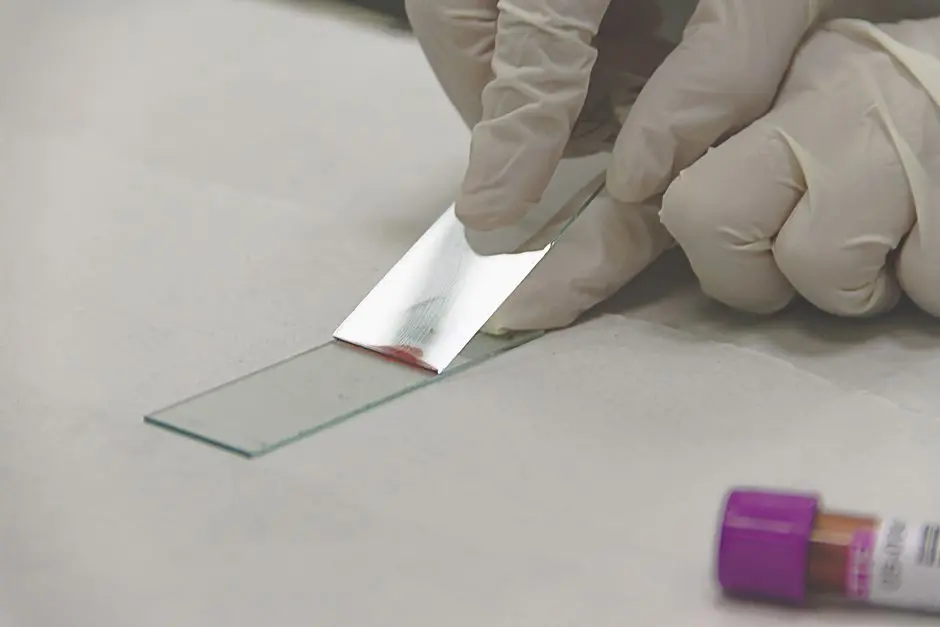
To truly master CLSM, we must proactively anticipate and circumvent common pitfalls. Our journey begins with sample preparation, a critical step often underestimated. Inadequate mounting media, air bubbles, or excessive thickness can severely compromise image quality. We advocate for mounting media with refractive indices matching the objective lens and cover glass (typically 1.518), and meticulous attention to ensuring samples are thin enough (typically <100 µm for optimal single-photon confocal) to minimize light scattering and absorption. Choosing appropriate fluorophores is another strategic decision; ensure their excitation and emission spectra are well-separated to avoid crosstalk, and that they possess high photostability to withstand laser illumination.
During image acquisition, balancing parameters is crucial. Laser power should be minimized to reduce photobleaching and phototoxicity, while still providing sufficient signal. Excessive detector gain can introduce noise, manifesting as grainy images, whereas insufficient gain can lead to weak signals. We rigorously calibrate these settings. The pinhole diameter directly impacts optical section thickness and signal intensity; a smaller pinhole yields thinner sections and higher resolution but reduces signal. A practical starting point is typically 1 Airy unit. Pixel dwell time (the duration the laser stays on each pixel) affects signal-to-noise ratio (SNR) and acquisition speed; longer dwell times improve SNR but increase photobleaching and total imaging time. Finally, proper image processing, including judicious use of deconvolution algorithms and 3D rendering software, can further enhance the quality and interpretability of your confocal data, but we always prioritize acquiring robust raw data.
Evolving Beyond: Advanced Confocal Techniques
Our journey into confocal microscopy extends beyond its foundational principles to embrace cutting-edge techniques that push the boundaries of biological imaging. One such advancement is Multi-Photon Microscopy (MPM). Instead of a single high-energy photon, MPM uses two or more lower-energy photons simultaneously to excite a fluorophore. This requires extremely high photon density, achieved by ultrashort pulsed infrared lasers. The key advantage is that excitation is highly localized to the focal plane, significantly reducing out-of-focus photobleaching and phototoxicity, enabling deeper tissue penetration (up to 1 mm) in scattering samples like intact brains. We deploy MPM for imaging in living organisms where depth and reduced damage are critical.
Another transformative development is the integration of confocal principles with Super-Resolution Microscopy (SRM) methods. While traditional confocal resolution is diffraction-limited, techniques like STimulated Emission Depletion (STED) microscopy directly extend the confocal concept. STED uses a second 'depletion' laser to de-excite fluorophores at the periphery of the excitation spot, effectively shrinking the fluorescent point spread function and achieving spatial resolutions down to ~20-50 nm, far surpassing the diffraction limit. Other SRM techniques, such as Structured Illumination Microscopy (SIM) and single-molecule localization microscopy (PALM/STORM), while not strictly confocal, often complement confocal data by providing molecular-level detail. These advanced modalities empower us to explore biological structures and dynamics at unprecedented nanometer scales, opening new frontiers in cell and molecular biology.
Strategic Deployment: Maximizing Your Confocal Investment
Maximizing the return on our confocal microscopy investment transcends mere technical operation; it demands strategic planning and meticulous execution. We commence with a robust experimental design. Clearly define your biological question, select appropriate fluorophores, and rigorously plan your controls. Consider the spatial and temporal resolution required for your specific research hypothesis. Are you tracking dynamics over minutes or hours? Do you need subcellular detail or broader tissue context? These decisions dictate your choice of objective, scanning speed, and acquisition parameters.
During data acquisition, consistent methodology is paramount. Maintain detailed records of all settings, from laser power and detector gain to pinhole size and pixel dimensions. This meticulous documentation is crucial for reproducibility and troubleshooting. For data interpretation, resist the urge to over-process. Prioritize honest representation of your raw data. Utilize appropriate software for 3D reconstruction and quantitative analysis, but always critically evaluate the impact of any digital manipulation. Regular microscope maintenance is non-negotiable; ensure objective lenses are clean, and regularly check laser alignment and detector calibration. Collaborate with core facility managers or experienced users to leverage their expertise. Finally, stay abreast of future trends. The field of microscopy is rapidly evolving, with ongoing advancements in detector sensitivity, faster scanning technologies (e.g., resonant scanners), and AI-driven image analysis. By embracing these strategic principles, we not only optimize our current confocal experiments but also position ourselves to harness the full potential of future innovations in biological imaging.
Key Takeaways
Core Principle: Optical Sectioning
Confocal microscopy precisely eliminates out-of-focus light using a pinhole aperture optically conjugate to the focal plane. This allows only light from the illuminated focal point to reach the detector, creating thin, high-contrast optical sections. By scanning point-by-point and collecting these sections, we construct accurate 3D representations.
Key Components for Precision
CLSM relies on integrated components: specific wavelength lasers for excitation, galvanometer scanners for precise X-Y movement, high numerical aperture objective lenses for light collection, dichroic mirrors for separating excitation/emission light, emission filters for signal specificity, a crucial pinhole for spatial filtering, and sensitive PMT detectors for signal conversion. Each element is vital for high-quality image acquisition.
Transformative Biological Applications
CLSM offers superior axial resolution, enabling detailed 3D reconstructions, reduced photobleaching/phototoxicity, and robust live-cell imaging capabilities. It is critical for studying protein localization, organelle dynamics, neuronal architecture, developmental processes, and various cellular interactions across diverse biological fields, offering insights unattainable with conventional microscopy.
Optimizing Acquisition: Best Practices & Pitfalls
Successful confocal imaging demands meticulous sample preparation (optimal mounting media, thickness), careful fluorophore selection (spectral separation, photostability), and precise parameter calibration. We minimize laser power, balance detector gain, set the pinhole to typically 1 Airy unit, and optimize pixel dwell time. Proper post-processing enhances data, but robust raw data acquisition is paramount to avoid artifacts and ensure interpretability.
Advanced Techniques: Deeper & Sharper Insights
Beyond standard CLSM, techniques like Multi-Photon Microscopy (MPM) allow deeper tissue penetration with minimal phototoxicity by using multiple low-energy photons. Super-Resolution Microscopy (SRM) methods such as STED microscopy push resolution beyond the diffraction limit (down to ~20-50 nm) by spatially confining fluorescence, offering unprecedented molecular-level detail and opening new avenues for biological discovery.
FAQ
-
How does confocal microscopy improve image resolution compared to widefield microscopy?
Confocal microscopy dramatically improves resolution, particularly in the Z-axis (depth), by employing a pinhole aperture. This pinhole acts as a spatial filter, blocking out-of-focus light emitted from areas above and below the focal plane. As a result, only light originating from the precisely defined focal volume reaches the detector, leading to sharper images with superior contrast and the ability to create clear optical sections, enabling high-fidelity 3D reconstructions.
-
What are the primary applications of CLSM in biological research?
CLSM is indispensable across numerous biological disciplines. Its primary applications include high-resolution 3D imaging of cells and tissues, precise localization of proteins and organelles, and the study of dynamic cellular processes through live-cell imaging. Researchers widely use it in neuroscience for neuronal morphology, developmental biology for tracking cell fate, cancer research for tumor microenvironment analysis, and microbiology for biofilm studies, among many others, due to its ability to minimize phototoxicity and photobleaching compared to traditional methods.
-
What is the 'Airy unit' in confocal microscopy, and why is it important?
The 'Airy unit' refers to the diameter of the Airy disk, which is the central bright spot of a diffraction pattern produced when light passes through a circular aperture, such as an objective lens. In confocal microscopy, the pinhole diameter is often expressed in Airy units. Setting the pinhole to 1 Airy unit typically provides the optimal balance between lateral resolution and signal intensity, ensuring effective out-of-focus light rejection while retaining sufficient light for detection. Adjusting the pinhole size directly influences the optical section thickness and the signal-to-noise ratio of your images.