
> Laboratory Techniques > Microscopy and Cellular Imaging > Uncover Optical Sectioning in Confocal Microscopy for Precise 3D Biology
Uncover Optical Sectioning in Confocal Microscopy for Precise 3D Biology
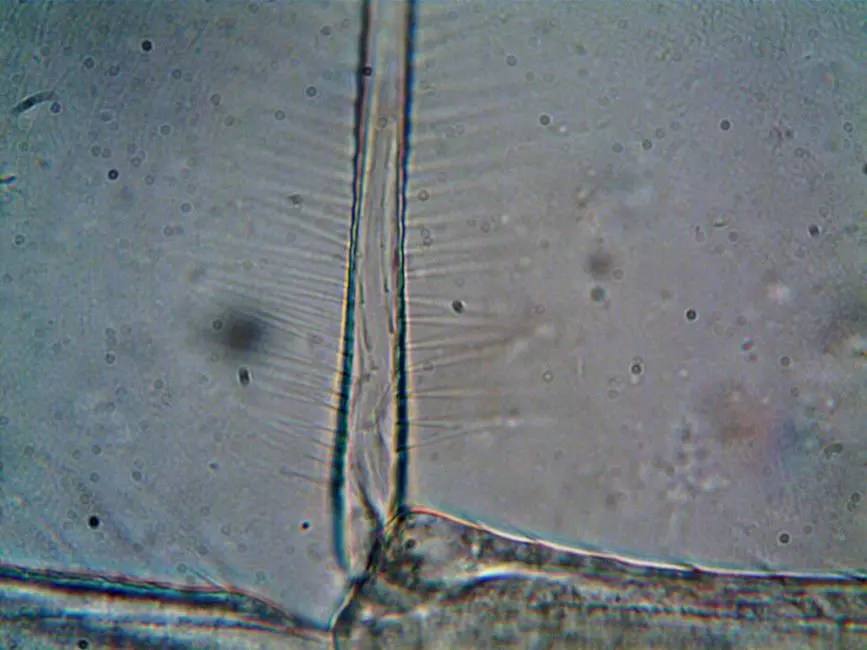
Envision a biological sample, alive and complex, yet obscured by the very light we use to observe it. Traditional widefield microscopy, while foundational, often delivers images muddled by out-of-focus light, rendering the three-dimensional intricacies of cells and tissues frustratingly indistinct. This fundamental limitation hinders our quest to decipher cellular mechanisms with precision.
We face a constant challenge: how to observe detailed structures deep within specimens without physical slicing. Confocal microscopy emerges as the strategic solution, ingeniously overcoming this optical hurdle by leveraging a powerful principle known as optical sectioning. This article meticulously dissects the core mechanics behind this revolutionary imaging technique, empowering us to capture pristine, high-resolution images in 3D. We forge a path to understanding how the confocal microscope precisely isolates light from a single focal plane, rejecting unwanted blur and unlocking unparalleled clarity. Prepare to elevate your understanding and master this critical capability, essential for advancing your research in advanced laboratory imaging methods for biological structures.
I. The Imperative for Optical Sectioning: Bridging the Gap from Widefield to Confocal
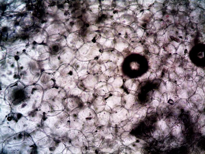
In our relentless pursuit of biological truth, the microscope stands as our primary gateway. Yet, conventional widefield fluorescence microscopy, despite its ubiquity, presents inherent limitations that can obscure critical biological insights. When illuminating an entire specimen simultaneously, light emanating from planes above and below the focal point inevitably reaches the detector. This out-of-focus light creates a pervasive haze, diminishing contrast, reducing resolution, and fundamentally preventing the clear visualization of discrete structures within a three-dimensional sample. Imagine trying to read text through a dense fog – that is the challenge widefield presents for thick specimens.
We demand clarity, depth, and the ability to reconstruct intricate 3D architectures without invasive physical sectioning. This is precisely where the concept of optical sectioning becomes not just beneficial, but absolutely imperative. Optical sectioning empowers us to virtually slice through a specimen, capturing discrete focal planes without causing physical damage or altering the biological context. This non-invasive approach preserves the sample's integrity, allowing for dynamic observations and precise volumetric reconstructions. It represents a paradigm shift from viewing a flattened projection to meticulously building a detailed, multi-layered representation of biological reality. The capacity to isolate fluorescence from a specific, thin optical slice unlocks unprecedented opportunities for analyzing cellular interactions, organelle dynamics, and tissue organization with surgical precision, thus accelerating our understanding of complex biological systems.
II. Unpacking the Mechanism: How Confocal Microscopy Achieves Optical Sectioning
At the heart of confocal microscopy’s transformative power lies its ingenious optical design, specifically engineered to achieve precise optical sectioning. The process begins with a focused laser beam, acting as a pinpoint light source, scanning across the specimen. Unlike widefield microscopy, which floods the entire field of view with light, the confocal system excites fluorophores within only a minute, diffraction-limited volume at the precise focal point. As the excited fluorophores emit light, this emitted fluorescence travels back through the objective lens.
The crucial differentiator, the cornerstone of optical sectioning, is the placement of a pinhole aperture in the conjugate focal plane – precisely aligned with the laser’s focal point. Only light originating from this specific focal plane passes through the pinhole to reach the detector. Critically, any light emitted from fluorophores above or below the focal plane – the out-of-focus light that plagues widefield imaging – is largely blocked by the opaque areas surrounding the pinhole. This sophisticated spatial filtering acts as an optical gate, effectively rejecting stray photons and dramatically enhancing image contrast and resolution within the selected focal slice. As the laser systematically scans across the x-y plane, and then the z-axis is incrementally adjusted, a series of optical sections are collected. These individual sections, devoid of out-of-focus blur, can then be computationally stacked to reconstruct a high-fidelity, three-dimensional representation of the specimen, truly revealing its internal architecture with unparalleled clarity.
III. Precision Engineering: Key Components and Their Role in Sectioning Quality
Optimizing optical sectioning in confocal microscopy demands a deep understanding of its integrated components, each playing a critical role in sculpting the final image quality. Let's dissect the vital players:
- Laser Light Source: We deploy highly stable, monochromatic lasers to provide the intense, specific excitation wavelengths necessary to activate fluorophores. The quality and stability of this light source directly impact signal intensity and consistency across the scan.
- Objective Lens: This is a paramount component. High numerical aperture (NA) objectives are non-negotiable for superior optical sectioning. A higher NA means a wider cone of light collection, leading to a tighter focal spot and, consequently, a thinner optical slice and enhanced lateral resolution. We consistently choose objectives with high NAs (e.g., 1.3 to 1.45 for oil immersion) for demanding 3D reconstructions.
- Dichroic Mirrors: These specialized filters selectively reflect excitation light towards the specimen while efficiently transmitting emitted fluorescence towards the detector. Their spectral properties are precisely matched to the fluorophores in use, ensuring optimal light separation and minimizing spectral crosstalk.
- Pinhole Aperture: The undisputed champion of optical sectioning. Its diameter is adjustable, and its optimal setting is a critical decision. A smaller pinhole rejects more out-of-focus light, yielding a thinner optical section and better axial resolution, but at the cost of reduced signal intensity. Conversely, a larger pinhole increases signal but thickens the optical slice, compromising sectioning fidelity. We must find the sweet spot, often aiming for 1 Airy unit for diffraction-limited resolution, or slightly larger for increased signal in very dim samples.
- Detectors (PMTs/HyDs): High-sensitivity photomultiplier tubes (PMTs) or hybrid detectors (HyDs) convert the faint fluorescence signals into electrical signals. Their quantum efficiency and low noise characteristics are vital for capturing weak signals emanating from deep within the specimen, especially when using small pinholes for ultra-thin sections.
Each component must operate in concert, meticulously aligned and optimized, to achieve robust and reliable optical sectioning, allowing us to faithfully reconstruct the intricate biological landscapes.
IV. Mastering Resolution and Depth: Parameters for Optimal Optical Sectioning
To truly master optical sectioning, we must command the parameters that govern resolution and depth penetration. These aren't just theoretical concepts; they are the levers we pull to extract maximum information from our samples. The goal: maximize signal-to-noise ratio (SNR) while maintaining desired resolution and minimizing phototoxicity.
- Pinhole Size (Airy Unit): As previously noted, the pinhole is central. A setting of 1 Airy unit typically provides the best compromise between axial resolution (thinness of the optical section) and signal collection efficiency. Going below 1 Airy unit yields diminishing returns in resolution gains but significantly reduces signal, increasing acquisition time and photobleaching risks. Conversely, setting the pinhole too large sacrifices the very benefit of optical sectioning. We must calibrate this carefully.
- Numerical Aperture (NA) of the Objective: A higher NA objective intrinsically yields a smaller point spread function (PSF) – the smallest point of light the microscope can resolve. A tighter PSF directly translates to thinner optical sections and improved lateral (x-y) resolution. This is a non-negotiable factor for high-resolution 3D imaging.
- Wavelength of Light: Shorter excitation and emission wavelengths generally lead to better resolution, as resolution is inversely proportional to wavelength. However, shorter wavelengths also penetrate less deeply into tissue and can cause more phototoxicity. We strategically select fluorophores with emission spectra that allow for deep penetration when necessary, balancing resolution with imaging depth.
- Pixel Dwell Time: This refers to the duration the laser spends at each pixel. Longer dwell times collect more photons, improving SNR, especially in dim samples or when using small pinholes. However, extended dwell times increase total acquisition time and photobleaching, demanding a careful balance for live-cell imaging.
- Scan Speed: Directly related to pixel dwell time. Faster scan speeds reduce acquisition time and photobleaching but decrease SNR. Slower speeds enhance SNR but risk photobleaching and motion artifacts in live samples.
- Laser Power: Higher laser power increases signal but drastically accelerates photobleaching and phototoxicity. We always use the minimum laser power necessary to achieve adequate signal, preserving sample vitality.
- Detector Gain and Offset: These electronic settings adjust the sensitivity and dynamic range of the detector. We optimize them to ensure the full dynamic range is utilized without saturating the detector or introducing excessive noise.
Effective management of these parameters is not merely a technical exercise; it is a strategic imperative to unlock the full potential of confocal imaging for robust biological discovery.
V. Advanced Strategies & Best Practices: Elevating Your Optical Sectioning Capabilities
Achieving truly exceptional optical sectioning goes beyond simply understanding the mechanics; it demands strategic implementation and a keen awareness of best practices. We must proactively address challenges to maximize data quality and biological relevance.
- Sample Preparation is Paramount: A perfectly prepared sample is the foundation. Clear, well-mounted specimens with minimal autofluorescence significantly improve SNR. Ensure proper refractive index matching between the sample, mounting medium, and immersion oil. Inconsistent refractive indices lead to spherical aberrations, which degrade the PSF and compromise optical sectioning depth and quality. For live-cell imaging, we prioritize physiological conditions to minimize stress and movement.
- Fluorophore Selection: Choose bright, photostable fluorophores with narrow emission spectra to minimize crosstalk and maximize signal efficiency. Consider genetically encoded fluorophores for live-cell tracking, carefully evaluating their brightness and photostability over extended imaging sessions.
- Minimizing Phototoxicity and Photobleaching: This is a critical challenge, especially for live-cell imaging. We employ strategies such as using the lowest possible laser power, shorter pixel dwell times, limiting the number of Z-stacks, and utilizing environmental control chambers. Advanced techniques like spinning disk confocal or two-photon microscopy offer reduced phototoxicity for specific applications, extending our imaging capabilities.
- Deconvolution and Image Processing: While confocal intrinsically rejects out-of-focus light, deconvolution algorithms can further refine the image. These computational methods mathematically remove remaining haze and reassign light to its correct spatial origin, leading to sharper images and even thinner effective optical sections, particularly valuable for fine detail reconstruction. We deploy these tools judiciously to enhance clarity without introducing artifacts.
- Calibration and Maintenance: Regular calibration of the microscope (e.g., laser alignment, objective parfocality, pinhole centration) is non-negotiable. Consistent maintenance ensures optimal performance, preventing subtle misalignments that can degrade optical sectioning efficiency and introduce imaging artifacts.
- Data Interpretation and 3D Reconstruction: Understand the limitations of your system. Optical sectioning data, once acquired, requires robust 3D reconstruction software to generate meaningful volumetric representations. We meticulously analyze individual sections and their collective stack, critically evaluating any potential artifacts or distortions introduced during acquisition or processing.
By integrating these advanced strategies and best practices, we transcend basic imaging, transforming confocal microscopy into a precision instrument for profound biological discovery.
Key Takeaways
Core Principle of Optical Sectioning
Confocal microscopy achieves optical sectioning by using a pinhole aperture in the detection pathway. This pinhole is precisely aligned with the focal plane of the laser, allowing only light originating from that specific plane to reach the detector. Light from out-of-focus planes is blocked, resulting in a thin, sharp optical slice of the specimen.
Key Components for Sectioning
Essential components include a focused laser source, a high Numerical Aperture (NA) objective lens, dichroic mirrors for spectral separation, and critically, an adjustable pinhole aperture. Each plays a role in creating a tight focal point and filtering out unwanted light.
Optimizing Resolution and Depth
Optimal optical sectioning relies on balancing several parameters: pinhole size (typically 1 Airy unit for best resolution), high NA objectives for tighter focus, appropriate laser power (minimal necessary), and pixel dwell time to maximize signal while minimizing photobleaching and phototoxicity. Wavelength also influences resolution and penetration depth.
Strategic Best Practices
To elevate results, prioritize meticulous sample preparation (refractive index matching, clear specimens), judicious fluorophore selection, vigilant mitigation of phototoxicity/photobleaching (low laser power, fast scanning), and the strategic use of deconvolution algorithms to further refine optical sections. Regular calibration and critical data interpretation are also vital.
FAQ
-
How does pinhole size directly impact optical sectioning?
The pinhole size is paramount for optical sectioning. A smaller pinhole rejects more out-of-focus light, resulting in a thinner optical section and improved axial (z-axis) resolution. This enhances the ability to discern fine details along the depth of the sample. However, reducing the pinhole size also reduces the total amount of light reaching the detector, leading to a weaker signal and potentially increased noise. Conversely, a larger pinhole collects more light, increasing signal strength, but it allows more out-of-focus light to pass, making the optical section thicker and compromising axial resolution. We always aim for a balance, often around 1 Airy unit, to optimize for both resolution and signal-to-noise ratio.
-
What is the primary advantage of optical sectioning in confocal microscopy over widefield microscopy?
The primary advantage is the ability to acquire high-resolution images from specific focal planes within a thick sample, virtually creating 'optical slices' without physically cutting the specimen. Widefield microscopy illuminates the entire sample and collects light from all depths simultaneously, resulting in blurred images due to out-of-focus light. Optical sectioning, through its pinhole mechanism, meticulously rejects this out-of-focus light, yielding sharp, high-contrast images of a single focal plane. This allows for precise 3D reconstruction of complex biological structures, detailed analysis of cellular components, and accurate quantification of fluorescence signals within specific cellular compartments, all while preserving the sample's integrity.
-
Can optical sectioning be performed on live biological samples, and what challenges does it present?
Yes, optical sectioning is extensively used for live biological samples, offering unparalleled insights into dynamic cellular processes in 3D. However, live-cell imaging presents unique challenges. The primary concerns are phototoxicity (damage to cells from prolonged laser exposure) and photobleaching (irreversible loss of fluorophore fluorescence). To mitigate these, we must minimize laser power, reduce pixel dwell times, optimize fluorophore selection for high brightness and photostability, and maintain strict environmental control (temperature, CO2, humidity) to ensure cell viability. Movement artifacts from living cells can also degrade image quality, requiring faster scan speeds or specialized image processing techniques.