
> Laboratory Techniques > Microscopy and Cellular Imaging > Reveal Life's Microcosm: Applications of Fluorescence Microscopy
Reveal Life's Microcosm: Applications of Fluorescence Microscopy
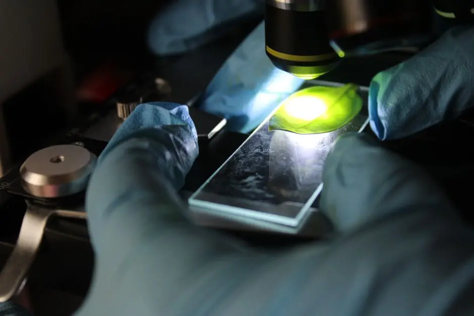
In the quest to decipher life's intricate dance, scientists relentlessly seek tools that pierce the veil of the microscopic. Fluorescence microscopy stands as a beacon in this endeavor, transforming our understanding of cellular architecture, molecular interactions, and dynamic biological processes. This powerful technique, leveraging the unique property of specific molecules to emit light after absorbing it, has revolutionized biology, enabling unprecedented visualization of life at its most fundamental level. We unlock the full spectrum of its utility, moving beyond mere observation to profound discovery.
This definitive guide meticulously explores the diverse applications of fluorescence microscopy, from unraveling the secrets of cellular organization to pinpointing disease mechanisms and advancing drug discovery. We delve into the core principles that empower this technology and illuminate how it serves as an indispensable instrument across myriad scientific disciplines. Prepare to optimize your understanding and harness the full potential of this revolutionary technique, solidifying its place among essential laboratory imaging methods for biological structures. We forge insights into its practical implementation, reveal common pitfalls, and uncover best practices that elevate research precision and impact. Join us as we dissect the methodologies that bring the invisible world to vibrant light.
Illuminating Principles: The Mechanism Behind Fluorescence
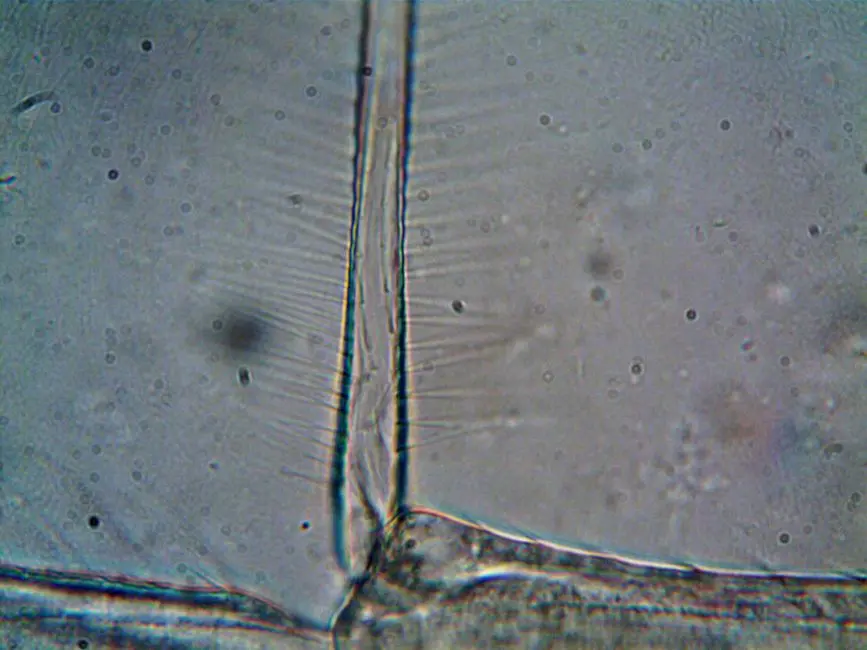
The core of fluorescence microscopy hinges on a precise photophysical process where specific molecules, known as fluorophores, absorb light at a particular wavelength (excitation) and subsequently emit light at a longer, lower-energy wavelength (emission). This phenomenon, characterized by the Stokes shift – the difference between the peak excitation and emission wavelengths – forms the basis of highly specific biological visualization. Unlike traditional brightfield microscopy, which relies on the absorption or scattering of light to create contrast, fluorescence microscopy actively generates its own signal from specific labels, allowing for the visualization of targets against a dark, often black, background. This dramatically enhances contrast and specificity.
We classify fluorophores into two main categories: intrinsic, such as certain amino acids or metabolites, and extrinsic, which are dyes or fluorescent proteins introduced into the sample. Genetically encoded fluorescent proteins, like GFP (Green Fluorescent Protein) and its myriad variants (e.g., mCherry, YFP), have revolutionized live-cell imaging due to their non-invasive nature. Synthetic organic dyes, such as DAPI for DNA, Alexa Fluors, or Cyanine dyes, offer a broad spectrum of colors and photostability, often used in fixed samples.
A typical fluorescence microscope comprises several essential components: a powerful light source (e.g., mercury lamp, LED, or laser) for excitation, a set of filters (excitation, emission, and dichroic mirror) to precisely select wavelengths, and a highly sensitive detector (e.g., CCD or sCMOS camera, or photomultiplier tube) to capture the emitted light. The dichroic mirror is particularly critical, reflecting excitation light towards the sample while allowing emitted fluorescence to pass through to the detector. The primary advantages of this technique are its exceptional specificity and high sensitivity. We can precisely target and visualize individual molecules, cellular structures, or even entire organisms with minimal background noise. However, we must always consider the potential for phototoxicity and photobleaching, ensuring our experimental parameters are optimized.
Charting the Cellular Landscape: Organelles, Proteins, and Dynamics
We employ fluorescence microscopy to precisely localize and visualize subcellular organelles, individual proteins, and complex cellular architectures. This capability is fundamental to understanding cellular function. Through techniques like immunofluorescence, where antibodies conjugated to fluorophores bind to specific cellular targets, and fluorescent protein tagging, we can illuminate structures such as mitochondria, endoplasmic reticulum, actin filaments, and microtubules with unprecedented clarity. The specificity of these labels ensures that only the intended structures or molecules are highlighted, reducing ambiguity in observations.
A pivotal application lies in live-cell imaging, allowing us to track dynamic processes in real-time. We observe cellular migration, protein trafficking pathways, membrane dynamics, and the intricate choreography of mitosis and meiosis. This real-time visualization provides critical insights into the kinetics and spatial regulation of biological events that are often obscured in fixed samples. For instance, we can follow the uptake of molecules via endocytosis or monitor the dynamic remodeling of the cytoskeleton in response to external stimuli.
Beyond qualitative observation, fluorescence microscopy enables quantitative analysis. We measure protein expression levels, analyze the spatial distribution of molecules within a cell, and quantify changes in cellular morphology over time. Good Practice: For live-cell imaging, we prioritize the use of genetically encoded fluorescent proteins (e.g., GFP variants). These fusions minimize perturbation to the cell and avoid fixation artifacts, providing a more accurate representation of biological processes in their native context. This approach is superior for dynamic studies where preserving cell viability and function is paramount. We optimize laser power and exposure times to balance signal detection with minimizing phototoxicity, a constant challenge in live-cell experiments.
Deciphering Molecular Dialogues: Interactions and Signaling
Fluorescence microscopy serves as an indispensable tool for unraveling the intricate dialogues between molecules and within signaling pathways. We utilize advanced techniques to investigate protein-protein interactions, monitor gene expression, and track physiological changes with remarkable precision. One powerful method is FRET (Förster Resonance Energy Transfer), which detects interactions between two fluorophores when they are in very close proximity (typically 1-10 nanometers). By labeling two interacting proteins with a donor and an acceptor fluorophore, we observe energy transfer from the excited donor to the acceptor, indicating a direct molecular association. This provides high spatial and temporal resolution for interaction studies within living cells.
Beyond FRET, we perform co-localization studies to assess whether two different molecules reside within the same cellular compartment or microdomain. While co-localization suggests potential interaction, it is crucial to recognize that it does not definitively prove direct binding. Common Error: Misinterpreting mere spatial overlap (co-localization) as a direct physical interaction. We leverage FRET or complementary biochemical assays to provide higher confidence for close proximity or direct binding.
Fluorescence microscopy also enables the imaging of reporter genes, where a fluorescent protein's expression is driven by a specific promoter. This allows us to monitor transcriptional activity and gene regulation in real-time, providing insights into cellular responses to various stimuli or developmental cues. Furthermore, the development of fluorescent biosensors – genetically encoded or synthetic probes that change their fluorescence properties in response to specific ions (e.g., calcium), pH, ATP, or enzyme activity – allows us to track dynamic physiological changes within cells and tissues. We apply these biosensors to study neural activity, metabolic shifts, and stress responses, mapping the spatio-temporal dynamics of cellular signaling networks.
From Bench to Bedside: Diagnostics and Therapeutic Discovery
Fluorescence microscopy drives innovation across medical diagnostics and drug discovery, bridging fundamental research with clinical applications. In disease diagnostics, we leverage its specificity for rapid and sensitive detection of pathogens. For instance, fluorescently labeled antibodies or nucleic acid probes can identify bacteria, viruses, or parasites directly in clinical samples, accelerating diagnosis and guiding treatment strategies. This is particularly critical in situations requiring quick identification of infectious agents.
In cancer research, fluorescence microscopy is indispensable. We visualize tumor margins during surgery, track metastatic cells in preclinical models, and identify specific biomarkers for diagnosis and prognosis. For example, fluorescent probes targeting specific cancer cell receptors can highlight malignant tissues, allowing for more precise surgical removal and reduced recurrence. In neuroscience, we map complex neuronal networks, study synaptic plasticity, and investigate the cellular pathology of neurodegenerative diseases such as Alzheimer's and Parkinson's. Fluorescent indicators can reveal calcium dynamics in neurons, offering insights into neural circuit function and dysfunction.
The impact extends significantly into drug discovery and development. High-throughput imaging (HTI) platforms, often leveraging automated fluorescence microscopy, allow us to screen vast libraries of compounds for their ability to modulate specific cellular pathways or interact with target proteins. This drastically accelerates the identification of potential therapeutic candidates. For example, HTI can screen >10<sup>5</sup> compounds per day, evaluating their effects on cell viability, protein translocation, or receptor activation. Moreover, we utilize fluorescence microscopy to study pharmacodynamics, monitoring drug uptake, distribution within cells and tissues, and their downstream cellular effects, providing crucial data for optimizing drug candidates and understanding their mechanisms of action. This comprehensive approach is vital for developing safer and more effective treatments.
Pushing Boundaries: Super-Resolution and Advanced Imaging
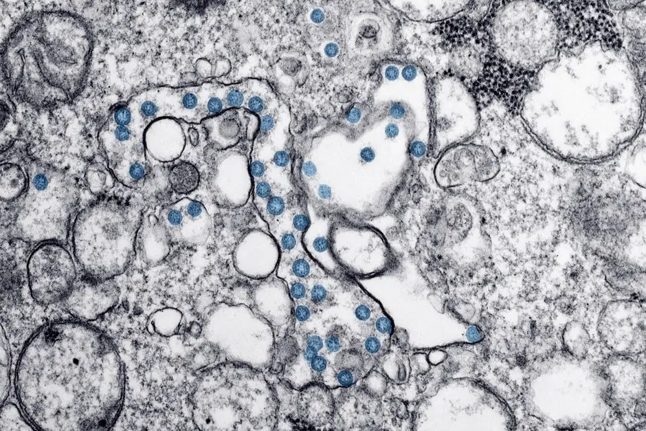
We continually push the boundaries of spatial and temporal resolution in fluorescence microscopy, unveiling biological phenomena at unprecedented scales. Advanced techniques overcome the traditional diffraction limit of light (approximately 200-250 nm), allowing us to visualize ultrastructural details previously only accessible with electron microscopy.
- FRAP (Fluorescence Recovery After Photobleaching): We employ FRAP to measure the mobility and diffusion rates of fluorescently labeled molecules within a living cell. By irreversibly photobleaching a small region and then observing the recovery of fluorescence as unbleached molecules diffuse into the area, we quantify molecular dynamics crucial for understanding protein turnover and membrane fluidity.
- FLIM (Fluorescence Lifetime Imaging Microscopy): FLIM provides information beyond mere intensity; it measures the decay rate of fluorescence, which is sensitive to the fluorophore's microenvironment. We utilize FLIM for applications such as quantifying FRET efficiency (indicating protein interactions) and sensing changes in cellular parameters like pH or oxygen concentration, offering functional insights alongside structural information.
- Super-Resolution Microscopy: This revolutionary suite of techniques shatters the diffraction barrier. STED (Stimulated Emission Depletion) microscopy uses a depletion laser to confine the fluorescence emission to a sub-diffraction volume. PALM (Photoactivated Localization Microscopy) and STORM (Stochastic Optical Reconstruction Microscopy) achieve super-resolution by localizing individual fluorescent molecules over time, reconstructing a high-resolution image from thousands of precise positions. These methods achieve resolutions significantly finer than <200</span> nm, revealing the nanoscale organization of organelles, protein clusters, and viral particles.
- Light-Sheet Microscopy: This technique employs a thin sheet of light to illuminate only the focal plane of the sample, minimizing phototoxicity and photobleaching outside this plane. We apply light-sheet microscopy for imaging large, delicate, and live samples (e.g., developing embryos, whole organs) over extended periods with minimal damage, providing critical insights into developmental biology and neurobiology.
Future Outlook: The integration of these advanced imaging modalities with artificial intelligence and machine learning is rapidly driving the field towards automated image analysis, pattern recognition, and quantitative biological modeling. We anticipate these synergies will further accelerate discovery by extracting deeper, more complex information from our data.
Mastering the Microscope: Best Practices and Troubleshooting
Achieving reliable and publishable data from fluorescence microscopy demands meticulous attention to experimental design and execution. We recognize that errors in sample preparation or data acquisition can lead to misleading results, undermining the scientific value of our efforts.
- Sample Preparation is Paramount: Whether we work with fixed or live cells, optimal sample preparation is critical. For fixed samples, this includes proper fixation (e.g., paraformaldehyde vs. methanol), gentle permeabilization, effective blocking to minimize non-specific antibody binding, and titrating antibody concentrations. Insider Tip: Always perform a titration for primary and secondary antibodies to determine the optimal concentration that yields a strong specific signal with minimal background fluorescence. This conserves valuable reagents and dramatically improves image quality and signal-to-noise ratio.
- Fluorophore Selection and Spectral Overlap: We carefully select fluorophores whose excitation and emission spectra are compatible with our microscope's filter sets and whose emissions are distinct enough to prevent spectral overlap (crosstalk) during multiplexed imaging. We utilize spectral unmixing algorithms when necessary to separate overlapping signals computationally.
- Mitigating Photobleaching and Phototoxicity: Photobleaching, the irreversible destruction of fluorophores by light, is a constant threat to live-cell imaging and prolonged acquisitions. We employ several strategies: reducing laser power, shortening exposure times, utilizing more photostable fluorophores, and adding anti-fade reagents to mounting media for fixed samples. For live cells, minimizing exposure to excitation light is paramount to reduce phototoxicity, which can alter cellular physiology.
- Addressing Autofluorescence: Biological samples often exhibit intrinsic fluorescence (autofluorescence), which can obscure specific signals. We identify sources of autofluorescence (e.g., collagen, elastin, NAD(P)H) and employ strategies like using red-shifted fluorophores, spectral unmixing, or chemical quenching to mitigate its impact.
- Robust Controls are Non-Negotiable: We always include appropriate controls to validate the specificity of our labeling and exclude artifacts. Examples include omitting the primary antibody, using isotype controls for immunofluorescence, imaging untransfected cells when using fluorescent protein fusions, and performing single-fluorophore controls for spectral unmixing.
- Rigorous Data Interpretation: Understanding the limitations of our imaging system and potential artifacts is crucial. We apply appropriate image processing techniques (e.g., deconvolution) and statistical rigor to our quantitative data, ensuring that our conclusions are robust and biologically meaningful.
Key Takeaways
Fundamental Principles and Components
Fluorescence microscopy operates on the principle of fluorophores absorbing light at one wavelength (excitation) and emitting it at a longer wavelength (emission). We utilize specific light sources, filters, and detectors to capture this emitted light, enabling high-contrast, specific visualization of biological targets. Fluorophores, whether intrinsic or engineered, are central to this technique, offering unparalleled specificity compared to brightfield.
Imaging Cellular Structures and Dynamics
We employ fluorescence microscopy to precisely localize and visualize subcellular organelles, individual proteins, and complex cellular architectures. Through techniques like immunofluorescence and fluorescent protein tagging, we track dynamic processes such as endocytosis, mitosis, and protein trafficking in real-time within live cells, providing crucial insights into cellular function and behavior.
Probing Molecular Interactions and Pathways
This technology is instrumental in deciphering molecular dialogues. We utilize FRET to detect nanometer-scale protein-protein interactions and employ fluorescent biosensors to monitor real-time changes in cellular microenvironments like pH or calcium levels. This allows us to map intricate signaling pathways and understand their regulation within living systems.
Impact on Disease and Drug Development
Fluorescence microscopy drives innovation in disease diagnostics, from identifying pathogenic microbes to visualizing tumor progression and metastasis. In drug discovery, we leverage high-throughput screening platforms based on fluorescence to rapidly evaluate compound efficacy, assess drug uptake, and monitor their pharmacological effects on target cells and pathways, accelerating therapeutic advancements.
Advancing Imaging Capabilities
We constantly push the boundaries of resolution and temporal imaging. Advanced techniques like FRAP measure molecular mobility, while FLIM provides environmental details. Super-resolution methods (STED, PALM/STORM) overcome the diffraction limit, revealing nanoscale structures. Light-sheet microscopy allows us to image large, delicate samples with minimal phototoxicity, opening new frontiers in developmental biology and neuroscience.
Optimizing Experimental Design and Execution
Mastering fluorescence microscopy demands rigorous attention to experimental detail. We prioritize meticulous sample preparation, careful fluorophore selection to avoid spectral overlap, and strategies to minimize photobleaching. Robust controls are indispensable for validating specificity and interpreting results accurately, ensuring the reliability and reproducibility of our biological discoveries.
FAQ
-
What are the primary advantages of fluorescence microscopy over traditional brightfield microscopy?
Fluorescence microscopy offers unparalleled specificity and sensitivity. We can selectively label and visualize specific molecules or structures within a complex cellular environment, providing high contrast against a dark background. This specificity allows for multiplexed imaging of multiple targets simultaneously and enables the study of dynamic processes in live cells, which is challenging with brightfield microscopy. -
What is the biggest challenge when performing fluorescence microscopy, and how can we mitigate it?
The most significant challenge is often photobleaching, the irreversible degradation of fluorophores due to light exposure, leading to signal loss. We mitigate this through several strategies: using lower excitation light intensity, shortening exposure times, incorporating anti-fade reagents into mounting media, and selecting more photostable fluorophores. Careful experimental design and optimization are crucial. -
How is fluorescence microscopy contributing to the future of medical diagnostics?
Fluorescence microscopy is pivotal for advanced medical diagnostics. We apply it for rapid and highly sensitive detection of pathogens, precise identification of disease biomarkers in tissues and cells, and even for guiding surgical procedures by visualizing tumor margins in real-time. Its ability to provide detailed molecular information at a cellular level empowers earlier and more accurate diagnoses, fostering personalized medicine. -
Can fluorescence microscopy be used to study processes in living organisms (in vivo)?
Absolutely. Fluorescence microscopy, particularly advanced forms like light-sheet microscopy or intravital microscopy, is extensively used for in vivo studies. We engineer cells or organisms to express fluorescent proteins, allowing us to track cellular migration, visualize neuronal activity, monitor tumor growth, or observe immune responses within a living context. This provides invaluable insights into complex biological processes that cannot be replicated in vitro.