
> Laboratory Techniques > Microscopy and Cellular Imaging > Uncover Cellular Ultrastructure: Electron Microscopy Unleashed
Uncover Cellular Ultrastructure: Electron Microscopy Unleashed
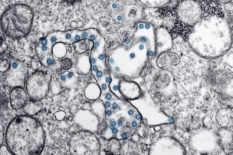
In the vast universe hidden within every cell, intricate machinery operates with breathtaking precision. Yet, deciphering these subcellular wonders demands vision far beyond the capabilities of conventional light microscopy. How do we pierce the veil of the nanoscale to comprehend the very architecture of life? We turn to electron microscopy (EM), a revolutionary technique that has fundamentally reshaped our understanding of biology.
This deep dive illuminates the transformative power of EM, detailing its principles, methodologies, and the unparalleled insights it delivers into cellular ultrastructure. We dissect the technical mastery required, from sample preparation to advanced imaging, providing an expert roadmap to interpreting these high-resolution revelations. Prepare to unlock the secrets of the cell, explore common pitfalls, and master the best practices that elevate EM from a mere tool to a true cornerstone among advanced laboratory imaging methodologies for biological structures. We forge a path to visual biological optimization, transforming invisible details into undeniable truths. Let us embark on this journey to dissect the cellular cosmos.
Conquer the Nanoscale: The Imperative of Electron Microscopy
Our quest to comprehend life's fundamental mechanisms often hinges on our ability to visualize its most minute components. While light microscopy serves as an invaluable gateway, its inherent physical limitations—dictated by the wavelength of visible light—constrain its resolution to approximately 200 nanometers. This barrier leaves the intricate world of cellular organelles, protein complexes, and viral structures largely opaque. To truly unravel the secrets of cellular function, we demand a deeper gaze, a tool capable of transcending this optical diffraction limit.
Electron microscopy emerges as our definitive answer, harnessing the wave-particle duality of electrons to achieve unparalleled resolving power. By accelerating electrons to extremely high velocities, we drastically reduce their associated de Broglie wavelength to mere picometers, orders of magnitude smaller than photons. This fundamental shift allows EM to resolve structures down to fractions of a nanometer, revealing cellular ultrastructure with exquisite detail—a capability impossible with traditional light methods. We do not merely observe cells; we penetrate their deepest architecture, mapping the endoplasmic reticulum's labyrinthine network, discerning the double membranes of mitochondria, and localizing ribosomes with unprecedented clarity. This leap in resolution is not just an incremental improvement; it is a paradigm shift, enabling us to correlate structure directly with function at the molecular level. We unlock insights critical for understanding disease mechanisms, drug targets, and basic cellular processes.
Architecting Clarity: Mastering Electron Microscopy Sample Preparation
The power of electron microscopy lies not only in the instrument itself but critically in the meticulous preparation of the biological specimen. Electrons, unlike photons, possess limited penetrating power and interact strongly with matter, necessitating ultra-thin samples and a vacuum environment. This process, often underestimated, is a true art form demanding precision and an intimate understanding of biological chemistry. We embark on a multi-stage journey to preserve cellular integrity, enhance contrast, and prepare samples for electron beam interaction.
The initial and paramount step involves fixation, rapidly preserving cellular structures in a near-native state. Glutaraldehyde cross-links proteins, followed by osmium tetroxide, which stabilizes lipids, provides contrast by binding to membranes, and acts as a secondary fixative. Inadequate fixation is a common error, leading to artifactual swelling or shrinkage. Next, dehydration gradually replaces water with organic solvents (e.g., ethanol, acetone), preparing the tissue for resin infiltration. This step must be carefully controlled to prevent structural damage. Subsequently, embedding involves infiltrating the tissue with a liquid plastic resin (e.g., epoxy resin) which is then polymerized to form a hard block. This rigidity enables ultra-thin sectioning. The final crucial step is ultramicrotomy, where sections typically 50-100 nm thick are cut using a diamond knife. These sections are then mounted on metal grids. Finally, post-staining with heavy metal salts like uranyl acetate and lead citrate enhances electron scattering, providing the necessary contrast to visualize cellular components. Each stage presents unique challenges, yet mastering them is non-negotiable for obtaining high-quality, artifact-free ultrastructural data. We ensure every detail is preserved, every structure revealed.
Dissecting the Invisible: Unveiling Ultrastructure with TEM and SEM
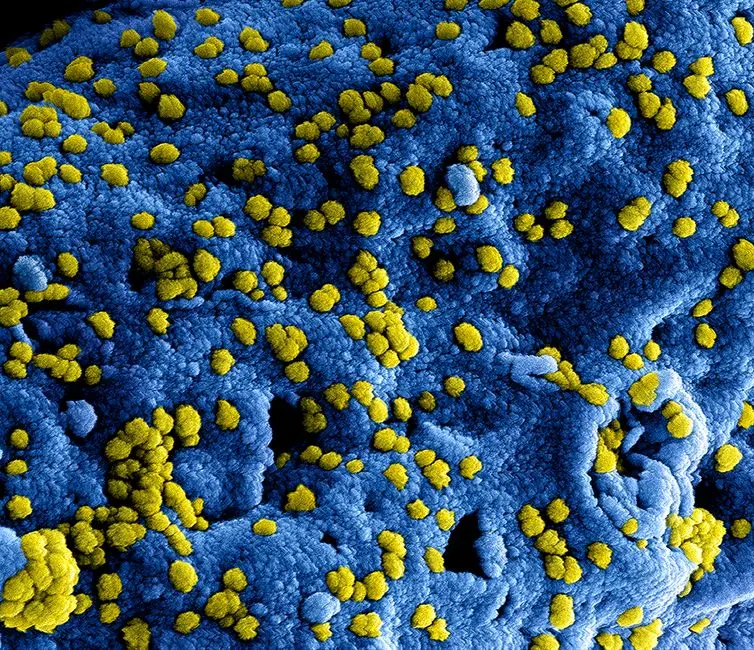
With expertly prepared samples, we deploy the two primary modalities of electron microscopy: Transmission Electron Microscopy (TEM) and Scanning Electron Microscopy (SEM). Each offers a distinct window into the cellular realm, providing complementary information about internal architecture and surface topography, respectively. Understanding their operational principles is key to interpreting the visual data they generate.
Transmission Electron Microscopy (TEM) operates much like a sophisticated slide projector. An electron beam passes through an ultra-thin specimen. Denser regions of the sample scatter more electrons, leading to fewer electrons reaching the detector, appearing dark in the final image. Less dense regions allow more electrons to pass through, appearing brighter. TEM excels at revealing internal cellular structures in two dimensions, allowing us to scrutinize the intricate folding of mitochondrial cristae, the precise arrangement of ribosomal subunits on the endoplasmic reticulum, the lumenal spaces within the Golgi apparatus, and the complex pore structures of the nuclear envelope. The resolution achieved permits the identification of individual protein complexes and even molecular arrangements. Scanning Electron Microscopy (SEM), conversely, focuses a finely tuned electron beam that scans across the specimen's surface. As the primary electrons strike the surface, they generate secondary electrons, backscattered electrons, and X-rays. Detectors collect these emitted signals, which are then used to construct a high-resolution, three-dimensional-like image of the specimen's surface morphology. SEM provides unparalleled views of cell surfaces, tissue organization, microbial biofilms, and extracellular matrix structures. While it offers less internal detail than TEM, its ability to render topographical information with great depth of field is invaluable. We leverage both techniques to construct a comprehensive, multi-dimensional understanding of cellular form and function.
Beyond Static Images: Dynamic Insights with Advanced EM Techniques
The capabilities of electron microscopy extend far beyond generating static, two-dimensional images. Modern advancements have transformed EM into a dynamic suite of tools, allowing us to probe molecular interactions, reconstruct three-dimensional cellular landscapes, and integrate data from across different imaging modalities. We explore these cutting-edge techniques that push the boundaries of ultrastructural analysis.
Cryo-Electron Microscopy (Cryo-EM) represents a revolutionary leap, earning a Nobel Prize for its impact. Instead of chemical fixation and dehydration, samples are flash-frozen in a thin layer of vitreous ice, preserving molecules and cellular components in their native, hydrated state. This avoids artifacts inherent in traditional preparation methods. Cryo-EM, particularly single-particle analysis, allows us to determine the near-atomic resolution structures of macromolecular complexes and membrane proteins, revealing their dynamic conformations. For visualizing cellular context, Electron Tomography offers a powerful solution. By acquiring a tilt series of TEM images—capturing the sample at multiple angles—and computationally reconstructing them, we generate high-resolution 3D models of specific cellular regions or organelles. This provides unprecedented insights into the spatial relationships and connectivity of internal structures. Furthermore, Immunogold Labeling permits the precise localization of specific proteins within the ultrastructural landscape. Antibodies tagged with electron-dense gold particles bind to target proteins, making their distribution visible under the EM. For a holistic view, Correlative Light and Electron Microscopy (CLEM) combines the functional insights of live-cell fluorescence microscopy with the ultrastructural detail of EM. We identify a dynamic event or specific protein localization in a live cell using light microscopy, then prepare that exact cell for EM to visualize the underlying ultrastructure responsible for that event. We integrate these powerful techniques to unravel the dynamic interplay within cells, propelling our understanding into new dimensions.
Strategic Deployment: Maximizing EM's Impact and Overcoming Challenges
Deploying electron microscopy effectively requires more than technical skill; it demands strategic planning, rigorous quality control, and an expert eye for interpretation. While EM offers unparalleled detail, it also presents challenges that, if not addressed, can compromise the validity of the findings. We outline key best practices and common pitfalls to ensure the integrity and impact of your ultrastructural studies.
Best Practices for Experimental Design: Clearly define your biological question to guide sample preparation and imaging strategy. Consider the required resolution and whether TEM, SEM, or advanced techniques like Cryo-EM or tomography are most appropriate. Implement rigorous controls to distinguish genuine biological structures from preparation artifacts. Image Acquisition and Analysis: Standardize imaging parameters, including magnification, voltage, and exposure times, for consistency across experiments. For quantitative analysis, employ systematic random sampling to ensure unbiased data collection. Utilize advanced image processing software for noise reduction, segmentation, and 3D reconstruction. Common Errors and Interpretation: A frequent pitfall is misinterpreting preparation artifacts—such as poor fixation, incomplete dehydration, or sectioning damage—as biological features. Always compare images to known ultrastructural standards and consult experienced microscopists. Over-interpretation of 2D sections as representative of 3D structures is another challenge; tomography directly addresses this. The dynamic nature of cells also means static EM images capture only a snapshot. Integrating EM with live-cell imaging via CLEM can mitigate this. We must cultivate a critical mindset, constantly questioning image origins and potential biases. By adhering to these principles, we elevate EM from a mere observation tool to a powerful engine of discovery, delivering robust and reproducible insights into the complexities of cellular life.
Key Takeaways
Electron Microscopy Fundamentals
Electron Microscopy (EM) provides unparalleled resolution by utilizing high-velocity electrons instead of photons, enabling visualization of cellular ultrastructure below the light microscope's diffraction limit. Its core strength lies in revealing intricate internal organelles and surface morphologies at sub-nanometer scales.
Critical Sample Preparation
Achieving high-quality EM images hinges on meticulous sample preparation: fixation (glutaraldehyde, osmium tetroxide), dehydration (ethanol, acetone), embedding (plastic resins), ultramicrotomy (50-100 nm sections), and heavy metal staining (uranyl acetate, lead citrate). Each step is crucial to preserve native structure and enhance contrast, with errors leading to significant artifacts.
TEM vs. SEM Distinction
Transmission Electron Microscopy (TEM) reveals internal cellular architecture by passing electrons through ultra-thin specimens, showing organelles in 2D. Scanning Electron Microscopy (SEM) provides high-resolution 3D-like images of specimen surfaces by scanning an electron beam and collecting scattered electrons.
Advanced EM Techniques
Modern EM includes: Cryo-EM for near-atomic resolution of macromolecules in native states; Electron Tomography for 3D reconstruction of cellular regions from tilt series; Immunogold Labeling for specific protein localization; and Correlative Light and Electron Microscopy (CLEM) for integrating dynamic functional data with ultrastructural detail.
Best Practices for Impact
Maximize EM's impact through: clear experimental design, rigorous controls, standardized image acquisition, and expert interpretation to differentiate biological features from common artifacts. A critical mindset and continuous learning are vital to extract reliable and profound insights into cellular biology.
FAQ
-
What is the primary advantage of electron microscopy over light microscopy?
The primary advantage of electron microscopy (EM) is its significantly higher resolution, achieved by using electrons instead of photons. Electrons have a much shorter wavelength, enabling EM to resolve structures down to fractions of a nanometer, whereas light microscopy is limited to approximately 200 nanometers. This allows EM to reveal the intricate ultrastructure of cells and organelles that are invisible under a light microscope.
-
Why is sample preparation so critical for electron microscopy?
Sample preparation is critical because electrons interact strongly with matter and require specific conditions for imaging. Biological samples must be: 1) fixed to preserve their native structure, 2) dehydrated as electrons cannot penetrate water, 3) embedded in resin for structural support, and 4) sectioned into ultra-thin slices (50-100 nm) for electron penetration. Improper preparation leads to artifacts, obscuring true cellular details and invalidating results.
-
What is the difference between TEM and SEM?
Transmission Electron Microscopy (TEM) passes an electron beam through an ultra-thin specimen to reveal internal cellular structures in high detail, much like an X-ray. Scanning Electron Microscopy (SEM) scans an electron beam across the surface of a specimen, generating an image from scattered electrons to reveal surface topography and three-dimensional morphology.
-
How does Cryo-EM overcome limitations of traditional EM?
Cryo-EM overcomes traditional EM limitations by flash-freezing samples in vitreous ice, preserving molecules and cellular structures in their native, hydrated state without the need for chemical fixation or dehydration. This minimizes artifacts and allows for the structural determination of macromolecules and complexes at near-atomic resolution, revealing their true dynamic conformations.
-
What are common artifacts to watch out for in EM images?
Common EM artifacts include:
- Poor fixation: Leads to cellular swelling, shrinkage, or membrane disruption.
- Incomplete dehydration: Results in patchy infiltration of resin.
- Sectioning damage: Appears as knife marks, compression, or chatter in the sections.
- Stain precipitates: Dark, irregular deposits often mistaken for cellular components.
- Vacuum damage: Can cause structural collapse if not properly embedded.
Careful attention to protocol and comparison with known healthy ultrastructure are essential for artifact identification.